. 2004 Dec;78(24):13954-65.
doi: 10.1128/JVI.78.24.13954-13965.2004.

Cyclin/CDK regulates the nucleocytoplasmic localization of the human papillomavirus E1 DNA helicase

Affiliations

Cyclin/CDK regulates the nucleocytoplasmic localization of the human papillomavirus E1 DNA helicase

Wentao Deng et al. J Virol. 2004 Dec.

Abstract

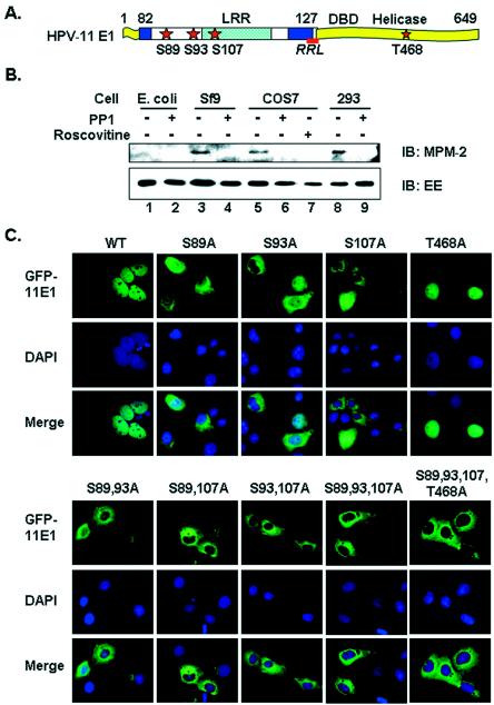
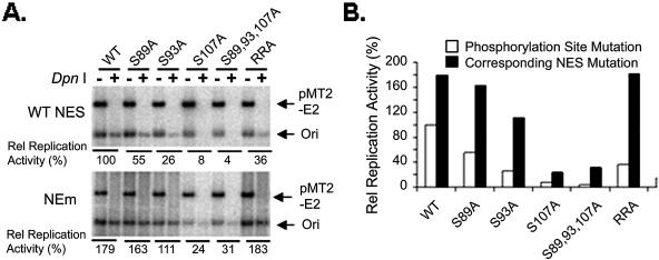
Cyclin-dependent kinases (CDKs) play key roles in eukaryotic DNA replication and cell cycle progression. Phosphorylation of components of the preinitiation complex activates replication and prevents reinitiation. One mechanism is mediated by nuclear export of critical proteins. Human papillomavirus (HPV) DNA replication requires cellular machinery in addition to the viral replicative DNA helicase E1 and origin recognition protein E2. E1 phosphorylation by cyclin/CDK is critical for efficient viral DNA replication. We now show that E1 is phosphorylated by CDKs in vivo and that phosphorylation regulates its nucleocytoplasmic localization. We identified a conserved regulatory region for localization which contains a dominant leucine-rich nuclear export sequence (NES), the previously defined cyclin binding motif, three serine residues that are CDK substrates, and a putative bipartite nuclear localization sequence. We show that E1 is exported from the nucleus by a CRM1-dependent mechanism unless the NES is inactivated by CDK phosphorylation. Replication activities of E1 phosphorylation site mutations are reduced and correlate inversely with their increased cytoplasmic localization. Nuclear localization and replication activities of most of these mutations are enhanced or restored by mutations in the NES. Collectively, our data demonstrate that CDK phosphorylation controls E1 nuclear localization to support viral DNA amplification. Thus, HPV adopts and adapts the cellular regulatory mechanism to complete its reproductive program.


Figures

FIG. 1.
FIG. 1.
In vivo phosphorylation and subcellular localization of wild-type and mutant forms of the HPV-11 E1 protein. (A) Functional domains and critical sequences regulating HPV-11 E1 nucleocytoplasmic localization. The sketch is not to scale. LRR, residues 82 to 127, was defined inthis work. DBD (DNA binding domain) and the helicase domain were defined previously. Stars, candidate CDK phosphorylation sites. Crosshatched box, a leucine-rich NES. Underlined segment, the consensus cyclin binding motif (RRL). Filled boxes, bipartite NLS. (B) In vivo phosphorylation of EE-E1. EE-E1 protein purified from different sources with (+) or without (−) prior treatment with PP1 was blotted with MPM-2 antibody, which recognizes phospho-S/T-P. The membrane was stripped and reprobed with anti-EE antibody. IB, immunoblot. (C) Subcellular localization of GFP-11E1 and phosphorylation site mutations in transfected COS7 cells. Upper panels show the green fluorescent signals for GFP-11E1, middle panels show the DAPI-stained nuclei, and the lower panels show the merged images. WT, wild type.
FIG. 2.
FIG. 2.
In vivo phosphorylation and subcellular localization of GFP-11E1 and mutations in the presence of inhibitors of CDKs or CRM1. Images were captured from transfected COS7 cells except those in panel C. (A) GFP-11E1 phosphorylation was blocked by mutations in the cyclin binding motif (RRL to RRA, KRA, or ARA). GFP-11E1 wild-type or mutated proteins were immunoprecipitated using polyclonal GFP antibody, blotted with MPM-2, and reprobed with monoclonal anti-GFP after stripping. (B) GFP-11E1 was localized to the cytoplasm (left panel) after CDK activity was inhibited by roscovitine (middle panels). In contrast, the nuclear localization of GFP-11E2 was not affected by roscovitine (right panels). Immunoprecipitation and Western blotting were performed as described above. (C) GFP-11E1 was localized to the cytoplasm after CDK activity was inhibited in p21-9 cells upon p21cip1 induction by IPTG (9) (left panels). The nuclear localization of GFP-11E2 was not affected by p21cip1 induction (right panels). (D) Inhibition of CRM1 with LMB (33, 53) restored nuclear localization of GFP-11E1 phosphorylation mutations. Twenty nanograms of LMB per milliliter was added to culture medium for 6 h. IB, immunoblot; WT, wild type; IP, immunoprecipitation.
FIG. 3.
FIG. 3.
Identification of the leucine-rich NES necessary and sufficient for HPV-11 E1 export. All images shown were taken from transfected COS7 cells. (A) The E1 proteins of HPVs contain a conserved LRR, which consists of the CDK phosphorylation sites (red letters), the cyclin binding motif (RRL, purple letters), a putative bipartite NLS (letters in italics), and a leucine-rich NES (green box). The consensus NES core sequence and the mutations in the consensus cyclin binding motif are illustrated above, and the NEm mutation is shown below. Underlined sequence, the leucine-rich NES core of 10 amino acids (residues 106 to 115). Green letters indicate the conserved large hydrophobic amino acids in the NES. Alignments with E1 sequences of other papillomaviruses are presented at the bottom. Note that the BPV-1 E1 lacks the NES. (B) Subcellular localization of GFP-11E1 mutated in the NES (NEm). WT, wild type. (C) Subcellular localization of EE-E1 and mutations, as revealed by indirect immunofluorescence (FITC) with anti-EE antibody. (D) Delineation of the functional NES in HPV-11 E1. E1 peptides were fused downstream of GFP. The positions of the appended peptides are indicated in parentheses.
FIG. 4.
FIG. 4.
Transient replication of wild-type and mutant forms of the GFP-11E1 protein. (A) Activities of the wild-type GFP-11E1, the phosphorylation mutations, and the corresponding NES mutations (NEm) in transient replication assays. Linearized ori (arrow) in DpnI lanes reflects the total amount of both the residual input ori and newly replicated ori plasmids, while DpnI+ lanes represent the newly replicated ori DNA. The signals from the pMT2-E2 plasmid served as a control for the complete DpnI digestion, equal ori plasmid input, transfection efficiency, and plasmid recovery. (B) Signals of replicated origin DNA (ori band from DpnI+ lane) were quantified, and the ratios to that obtained with wild-type E1 (100%) were presented as relative replication activities. WT, wild type.

Similar articles

Cited by

References

    1. Abraham, R. T. 2001. Cell cycle checkpoint signaling through the ATM and ATR kinases. Genes Dev. 15:2177-2196. - PubMed
    1. Amin, A. A., S. Titolo, A. Pelletier, D. Fink, M. G. Cordingley, and J. Archambault. 2000. Identification of domains of the HPV11 E1 protein required for DNA replication in vitro. Virology 272:137-150. - PubMed
    1. Auster, A. S., and L. Joshua-Tor. 2004. The DNA-binding domain of human papillomavirus type 18 E1: crystal structure, dimerization, and DNA binding. J. Biol. Chem. 279:3733-3742. - PubMed
    1. Bain, J., H. McLauchlan, M. Elliott, and P. Cohen. 2003. The specificities of protein kinase inhibitors: an update. Biochem. J. 371:199-204. - PMC - PubMed
    1. Bell, S. P., and A. Dutta. 2002. DNA replication in eukaryotic cells. Annu. Rev. Biochem. 71:333-374. - PubMed

Publication types

MeSH terms

LinkOut - more resources