. 2004 Dec;78(24):14003-11.
doi: 10.1128/JVI.78.24.14003-14011.2004.

Human metapneumovirus persists in BALB/c mice despite the presence of neutralizing antibodies

Affiliations

Human metapneumovirus persists in BALB/c mice despite the presence of neutralizing antibodies

Rene Alvarez et al. J Virol. 2004 Dec.

Abstract

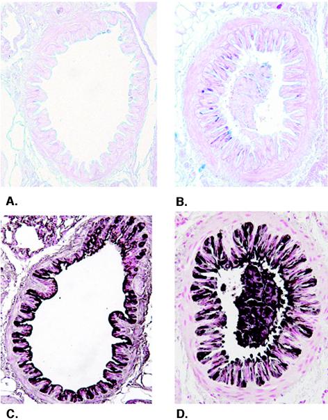
Human metapneumovirus (HMPV) has emerged as an important human respiratory pathogen causing upper and lower respiratory tract infections in young children and older adults. Recent epidemiological evidence indicates that HMPV may cocirculate with respiratory syncytial virus, and HMPV infection has been associated with other respiratory diseases. In this study, we show that BALB/c mice are susceptible to HMPV infection, the virus replicates in the lungs with biphasic growth kinetics in which peak titers occur at days 7 and 14 postinfection (p.i.), and infectious HMPV can be recovered from lungs up to day 60 p.i. In addition, we show that genomic HMPV RNA can be detected in the lungs for >/=180 days p.i. by reverse transcription-PCR; however, neither HMPV RNA nor infectious virus can be detected in serum, spleen, kidneys, heart, trachea, and brain tissue. Lung histopathology revealed prevalent mononuclear cell infiltration in the interstitium beginning at day 2 p.i. and peaking at day 4 p.i. which decreased by day 14 p.i. and was associated with airway remodeling. Increased mucus production evident at day 2 p.i. was concordant with increased bronchial and bronchiolar inflammation. HMPV-specific antibodies were detected by day 14 p.i., neutralizing antibody titers reached >/=6.46 log(2) end-point titers by day 28 p.i., and depletion of T cells or NK cells resulted in increased HMPV titers in the lungs, suggesting some immune control of viral persistence. This study shows that BALB/c mice are amenable for HMPV studies and indicates that HMPV persists as infectious virus in the lungs of normal mice for several weeks postinfection.


Figures

FIG. 1.
FIG. 1.
HMPV infection is associated with weight loss. BALB/c mice were intranasally infected with 106 PFU of HMPV. Mice were monitored for signs of clinical illness including weight loss, huddling, ruffled fur, and decreased mobility. (A) Mice were weighed, and the lungs were harvested to determine HMPV viral titers. Each time point represents the mean titer per gram of lung tissue ± SD from three experiments using three mice per experiment. (B) Total RNA was isolated from HMPV-infected BALB/c mouse lungs and assayed by RT-PCR with HMPV M gene-specific primers (top) or GAPDH primers (bottom) as an internal control. Lane 1, 1-kb molecular size marker; lane 2, day 0 p.i.; lane 3, day 7 p.i.; lane 4, day 14 p.i.; lane 5, day 28 p.i.; lane 6, day 60 p.i.; lane 7, day 90 p.i.; lane 8, day 150 p.i.; lane 9, day 180 p.i.; lane 10, 1-kb molecular size marker.
FIG. 1.
FIG. 1.
HMPV infection is associated with weight loss. BALB/c mice were intranasally infected with 106 PFU of HMPV. Mice were monitored for signs of clinical illness including weight loss, huddling, ruffled fur, and decreased mobility. (A) Mice were weighed, and the lungs were harvested to determine HMPV viral titers. Each time point represents the mean titer per gram of lung tissue ± SD from three experiments using three mice per experiment. (B) Total RNA was isolated from HMPV-infected BALB/c mouse lungs and assayed by RT-PCR with HMPV M gene-specific primers (top) or GAPDH primers (bottom) as an internal control. Lane 1, 1-kb molecular size marker; lane 2, day 0 p.i.; lane 3, day 7 p.i.; lane 4, day 14 p.i.; lane 5, day 28 p.i.; lane 6, day 60 p.i.; lane 7, day 90 p.i.; lane 8, day 150 p.i.; lane 9, day 180 p.i.; lane 10, 1-kb molecular size marker.
FIG. 2.
FIG. 2.
HMPV-specific antibody response. HMPV-specific antibodies were quantitated by ELISA following intranasal infection of BALB/c mice. Each time point represents the mean optical density (OD) ± SD from three separate experiments using three mice per experiment.
FIG. 3.
FIG. 3.
Histopathology of HMPV infection in mice. Interstitial inflammatory cell infiltrates were examined in the lung at (A) day 0, (B) day 2, (C) day 4, (D) day 7, (E) day 10, or (F) day 14 following hematoxylin and eosin staining. Magnification, ×25 (A to F).
FIG. 4.
FIG. 4.
Airway epithelial changes following HMPV infection. Levels of mucus production (A and B) and the airway epithelial cell marker CCSP (C and D) were determined in lung sections from mice at day 0 (A and C) and day 2 (B and D) after HMPV infection. Mucus production (pink to purple staining) is apparent in the airways of mice. CCSP staining (brown to black staining) is associated with altered airway epithelial morphology and staining of cell debris in the airway lumen (D). Magnification, ×300.
FIG. 5.
FIG. 5.
Antibody depletion of T cells or NK cells affects HMPV replication. BALB/c mice were depleted of T cells or NK cells prior to HMPV infection (106 PFU), and the lung virus titers were determined at day 7 p.i., day 28 p.i., or day 60 p.i. Data are presented as the median virus titers ± SD from three experiments using three mice per experiment.

Similar articles

Cited by

References

    1. Ahmed, R., L. A. Morrison, and D. M. Knipe. 1996. Persistence of viruses, p. 219-249. In B. N. Fields, D. M. Knipe, P. M. Howley, R. M. Chanock, J. L. Melnick, T. P. Monath, B. Roizman, and S. E. Straus (ed.), Fields virology, 3rd ed., vol. 1. Lippincott Williams & Wilkins, Philadelphia, Pa.
    1. Alvarez, R., L. P. Jones, B. S. Seal, D. R. Kapczynski, and R. A. Tripp. 2004. Serological cross-reactivity of members of the Metapneumovirus genus. Virus Res. 105:67-73. - PubMed
    1. Bastien, N., S. Normand, T. Taylor, D. Ward, T. C. Peret, G. Boivin, L. J. Anderson, and Y. Li. 2003. Sequence analysis of the N, P, M and F genes of Canadian human metapneumovirus strains. Virus Res. 93:51-62. - PMC - PubMed
    1. Belshe, R. B., L. S. Richardson, W. T. London, D. L. Sly, J. H. Lorfeld, E. Camargo, D. A. Prevar, and R. M. Chanock. 1977. Experimental respiratory syncytial virus infection of four species of primates. J. Med. Virol. 1:157-162. - PubMed
    1. Boivin, G., Y. Abed, G. Pelletier, L. Ruel, D. Moisan, S. Côté, T. C. Peret, D. D. Erdman, and L. J. Anderson. 2002. Virological features and clinical manifestations associated with human metapneumovirus: a new paramyxovirus responsible for acute respiratory-tract infections in all age groups. J. Infect. Dis. 186:1330-1334. - PubMed

Publication types

MeSH terms

LinkOut - more resources