. 2004 Dec;78(24):14057-61.
doi: 10.1128/JVI.78.24.14057-14061.2004.

Genetic screen for monitoring severe acute respiratory syndrome coronavirus 3C-like protease

Affiliations

Genetic screen for monitoring severe acute respiratory syndrome coronavirus 3C-like protease

Mariona Parera et al. J Virol. 2004 Dec.

Abstract

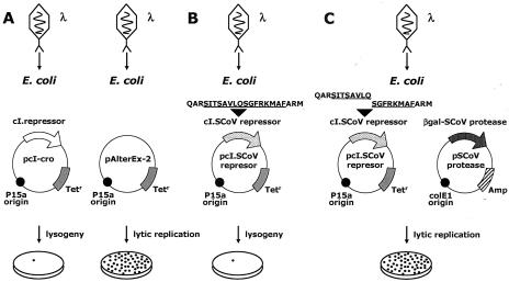
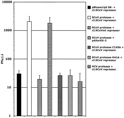
A novel coronavirus (SCoV) is the etiological agent of severe acute respiratory syndrome. Site-specific proteolysis plays a critical role in regulating a number of cellular and viral processes. Since the main protease of SCoV, also termed 3C-like protease, is an attractive target for drug therapy, we have developed a safe, simple, and rapid genetic screen assay to monitor the activity of the SCoV 3C-like protease. This genetic system is based on the bacteriophage lambda regulatory circuit, in which the viral repressor cI is specifically cleaved to initiate the lysogenic-to-lytic switch. A specific target for the SCoV 3C-like protease, P1/P2 (SAVLQ/SGFRK), was inserted into the lambda phage cI repressor. The target specificity of the SCoV P1/P2 repressor was evaluated by coexpression of this repressor with a chemically synthesized SCoV 3C-like protease gene construct. Upon infection of Escherichia coli cells containing the two plasmids encoding the cI. SCoV P1/P2-cro and the beta-galactosidase-SCoV 3C-like protease constructs, lambda phage replicated up to 2,000-fold more efficiently than in cells that did not express the SCoV 3C-like protease. This simple and highly specific assay can be used to monitor the activity of the SCoV 3C-like protease, and it has the potential to be used for screening specific inhibitors.


Figures

FIG. 1.
FIG. 1.
Amino acid sequence of the SCoV 3C-like protease engineered in the present study. The autocleavage sites of the protease are marked with vertical arrows above the sequences. The cleavage site used as a target site in the genetic screen described here is shaded. Underlined are the catalytic-site residues Cys145 and His41.
FIG. 2.
FIG. 2.
Lambda-based genetic screen to monitor the activity of SCoV 3C-like protease. This genetic screen system is based on the bacteriophage lambda cI-cro regulatory circuit, where the viral repressor cI is specifically cleaved to initiate the lysogenic-to-lytic switch. (A) Expression of the phage-encoded repressor (cI) results in repression of the bacteriophage's lytic functions (lysogeny). (B) SCoV target repressor containing the P1/P2 cleavage site; as illustrated in Fig. 3 and 4, this repressor efficiently represses the infecting phage (lysogeny). (C) When phages infect E. coli cells that express recombinant cI.SCoV repressor and β-Gal-SCoV 3C-like protease, infection results in lytic replication.
FIG. 3.
FIG. 3.
Selective growth of λ in E. coli cells coexpressing the β-Gal-SCoV 3C-like protease construct and the cI.SCoV repressor. Expression of the protease was induced with IPTG for 1 h, and the cells were infected with λ for three additional hours. The graph illustrates the resulting phage titer per microliter. Plasmids pBSK− and pAlterEX-2 were used as controls for the β-Gal-SCoV 3C-like protease construct and the cI.SCoV repressor, respectively. cI.SCoVmt was also used as a negative control for the cI.SCoV repressor. As shown, selection in cells coexpressing the β-Gal-SCoV 3C-like protease construct and the cI.SCoV3 repressor resulted in λ replication, whereas the replication of λ was severely compromised in cells expressing the mutant cI.SCoVmt repressor. Lack of phage replication was also observed in cells expressing mutated forms of β-Gal-SCoV 3C-like protease that included catalytic-site residue substitutions C145A and H41A. Similarly, expression of another protease (HCV serine protease) also prevented phage replication. Values are the means ± standard deviations (error bars) of at least four experiments.
FIG. 4.
FIG. 4.
The SCoV 3C-like protease reduces the expression levels of cI.CoV. The cI.SCoV and cI.SCoVmt (lane 4) repressors were coexpressed with the SCoV 3C-like protease. Expression of the protease was induced with IPTG for 3 h. The ODs of the cultures after 3 h (in the presence of IPTG) were measured to assure that equivalent amounts of total cell protein were blotted. No significant differences were observed when the ODs of the different cultures were compared, suggesting that the expression of the SCoV 3C-like protease did not affect the growth of the bacteria. Control proteases with catalytic residue substitutions C145A and H41A and another protease (HCV serine protease) were also included in this experiment (lanes 5, 6, and 8, respectively). Lane 7 cells were grown in the absence of IPTG. Reduced signal and cleavage products were observed only when the wild-type (wt) SCoV 3C-like protease was expressed (lane 1); cleavage products were also observed in the absence of IPTG, suggesting residual expression of the wild-type SCoV 3C-like protease (lane 7). E. coli JM109 cells were cotransformed with pAlterEx-2 repressor plasmids and the pBSK− plasmid containing wild-type or mutated SCoV 3C-like proteases. Cultures were lysed in sodium dodecyl sulfate (SDS)-polyacrylamide gel electrophoresis sample buffer, resolved in 18% gradient SDS-polyacrylamide gels, transferred to nitrocellulose membranes, and blocked in phosphate-buffered saline-0.1% Tween 20-10% nonfat dry milk. For immunochemical detection of the cI.SCoV repressor, membranes were subsequently incubated with rabbit serum containing polyclonal anti-cI antibodies (anti-cI sera; Invitrogen). Bound antibodies were visualized with peroxidase-linked anti-rabbit immunoglobulin G (Pierce) and the ECL Plus kit (Amersham Biosciences).

Similar articles

Cited by

References

    1. Anand, K., G. J. Palm, J. R. Mesters, S. G. Siddell, J. Ziebuhr, and R. Hilgenfeld. 2002. Structure of coronavirus main proteinase reveals combination of a chymotrypsin fold with an extra alpha-helical domain. EMBO J. 21:3213-3224. - PMC - PubMed
    1. Anand, K., J. Ziebuhr, P. Wadhwani, J. R. Mesters, and R. Hilgenfeld. 2003. Coronavirus main proteinase (3CLpro) structure: basis for design of anti-SARS drugs. Science 300:1763-1767. - PubMed
    1. Cabana, M., G. Fernandez, M. Parera, B. Clotet, and M. A. Martinez. 2002. Catalytic efficiency and phenotype of HIV-1 proteases encoding single critical resistance substitutions. Virology 300:71-78. - PubMed
    1. Cello, J., A. V. Paul, and E. Wimmer. 2002. Chemical synthesis of poliovirus cDNA: generation of infectious virus in the absence of natural template. Science 297:1016-1018. - PubMed
    1. Drosten, C., S. Gunther, W. Preiser, S. van der Werf, H. R. Brodt, S. Becker, H. Rabenau, M. Panning, L. Kolesnikova, R. A. Fouchier, A. Berger, A. M. Burguiere, J. Cinatl, M. Eickmann, N. Escriou, K. Grywna, S. Kramme, J. C. Manuguerra, S. Muller, V. Rickerts, M. Sturmer, S. Vieth, H. D. Klenk, A. D. Osterhaus, H. Schmitz, and H. W. Doerr. 2003. Identification of a novel coronavirus in patients with severe acute respiratory syndrome. N. Engl. J. Med. 348:1967-1976. - PubMed

Publication types

MeSH terms

Substances

Associated data

LinkOut - more resources