

Induction of deltaFosB in reward-related brain structures after chronic stress
- PMID: 15564575
- PMCID: PMC6730117
- DOI: 10.1523/JNEUROSCI.2542-04.2004
Induction of deltaFosB in reward-related brain structures after chronic stress
Abstract
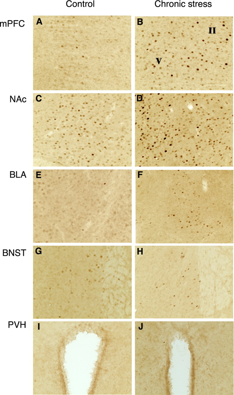
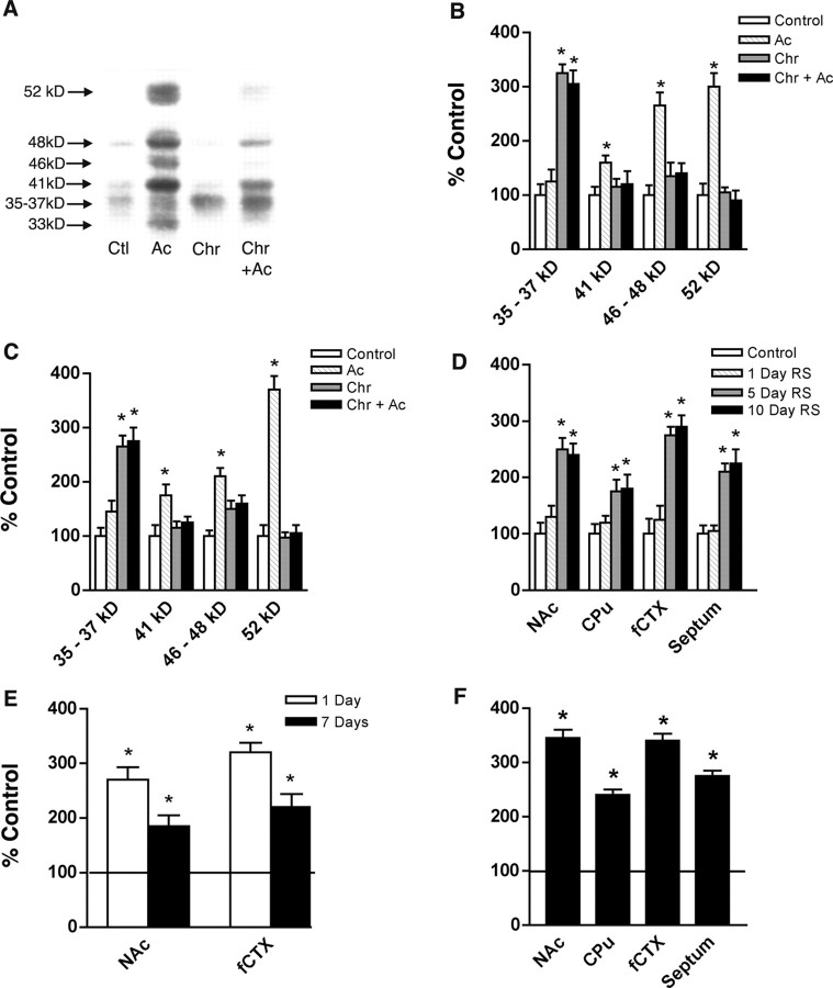
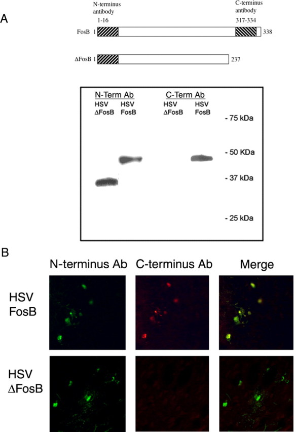
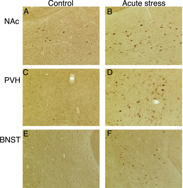
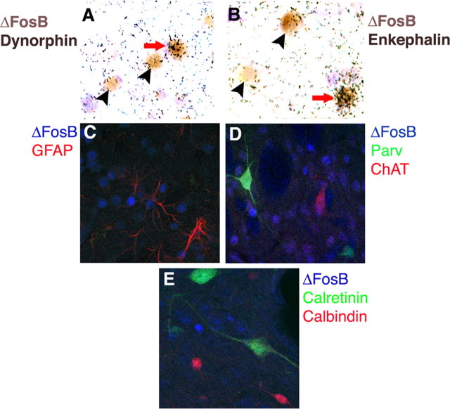
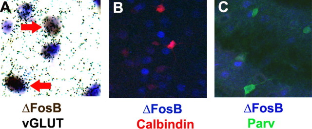
Acute and chronic stress differentially regulate immediate-early gene (IEG) expression in the brain. Although acute stress induces c-Fos and FosB, repeated exposure to stress desensitizes the c-Fos response, but FosB-like immunoreactivity remains high. Several other treatments differentially regulate IEG expression in a similar manner after acute versus chronic exposure. The form of FosB that persists after these chronic treatments has been identified as DeltaFosB, a splice variant of the fosB gene. This study was designed to determine whether the FosB form induced after chronic stress is also DeltaFosB and to map the brain regions and identify the cell populations that exhibit this effect. Western blotting, using an antibody that recognizes all Fos family members, revealed that acute restraint stress caused robust induction of c-Fos and full-length FosB, as well as a small induction of DeltaFosB, in the frontal cortex (fCTX) and nucleus accumbens (NAc). The induction of c-Fos (and to some extent full-length FosB) was desensitized after 10 d of restraint stress, at which point levels of DeltaFosB were high. A similar pattern was observed after chronic unpredictable stress. By use of immunohistochemistry, we found that chronic restraint stress induced DeltaFosB expression predominantly in the fCTX, NAc, and basolateral amygdala, with lower levels of induction seen elsewhere. These findings establish that chronic stress induces DeltaFosB in several discrete regions of the brain. Such induction could contribute to the long-term effects of stress on the brain.
Figures

Similar articles
-
Glucocorticoids regulation of FosB/ΔFosB expression induced by chronic opiate exposure in the brain stress system.PLoS One. 2012;7(11):e50264. doi: 10.1371/journal.pone.0050264. Epub 2012 Nov 21. PLoS One. 2012. PMID: 23185589 Free PMC article.
-
Induction of FosB/DeltaFosB in the brain stress system-related structures during morphine dependence and withdrawal.J Neurochem. 2010 Jul;114(2):475-87. doi: 10.1111/j.1471-4159.2010.06765.x. Epub 2010 Apr 23. J Neurochem. 2010. PMID: 20438612
-
Region-specific induction of deltaFosB by repeated administration of typical versus atypical antipsychotic drugs.Synapse. 1999 Aug;33(2):118-28. doi: 10.1002/(SICI)1098-2396(199908)33:2<118::AID-SYN2>3.0.CO;2-L. Synapse. 1999. PMID: 10400890
-
∆FosB: a transcriptional regulator of stress and antidepressant responses.Eur J Pharmacol. 2015 Apr 15;753:66-72. doi: 10.1016/j.ejphar.2014.10.034. Epub 2014 Nov 7. Eur J Pharmacol. 2015. PMID: 25446562 Free PMC article. Review.
-
DeltaFosB: a sustained molecular switch for addiction.Proc Natl Acad Sci U S A. 2001 Sep 25;98(20):11042-6. doi: 10.1073/pnas.191352698. Proc Natl Acad Sci U S A. 2001. PMID: 11572966 Free PMC article. Review.
Cited by
-
The Neurocircuitry of Posttraumatic Stress Disorder and Major Depression: Insights Into Overlapping and Distinct Circuit Dysfunction-A Tribute to Ron Duman.Biol Psychiatry. 2021 Jul 15;90(2):109-117. doi: 10.1016/j.biopsych.2021.04.009. Epub 2021 Apr 24. Biol Psychiatry. 2021. PMID: 34052037 Free PMC article. Review.
-
Neurological correlates of brain reward circuitry linked to opioid use disorder (OUD): Do homo sapiens acquire or have a reward deficiency syndrome?J Neurol Sci. 2020 Nov 15;418:117137. doi: 10.1016/j.jns.2020.117137. Epub 2020 Sep 15. J Neurol Sci. 2020. PMID: 32957037 Free PMC article. Review.
-
Daily limited access to sweetened drink attenuates hypothalamic-pituitary-adrenocortical axis stress responses.Endocrinology. 2007 Apr;148(4):1823-34. doi: 10.1210/en.2006-1241. Epub 2007 Jan 4. Endocrinology. 2007. PMID: 17204558 Free PMC article.
-
Striatal regulation of ΔFosB, FosB, and cFos during cocaine self-administration and withdrawal.J Neurochem. 2010 Oct;115(1):112-22. doi: 10.1111/j.1471-4159.2010.06907.x. Epub 2010 Aug 3. J Neurochem. 2010. PMID: 20633205 Free PMC article.
-
Modulation by Estradiol of L-Dopa-Induced Dyskinesia in a Rat Model of Post-Menopausal Hemiparkinsonism.Life (Basel). 2022 Apr 26;12(5):640. doi: 10.3390/life12050640. Life (Basel). 2022. PMID: 35629308 Free PMC article.
References
-
- Alibhai IN, Green TA, Nestler EJ (2004) In vivo and in vitro regulation of FosB mRNA isoforms. Soc Neurosci Abstr 30: 692.1.
-
- Atkins JB, Chlan-Fourney J, Nye HE, Hiroi N, Carlezon Jr WA, Nestler EJ (1999) Region-specific induction of deltaFosB by repeated administration of typical versus atypical antipsychotic drugs. Synapse 33: 118-128. - PubMed
-
- Bruijnzeel AW, Stam R, Compaan JC, Croiset G, Akkermans LM, Olivier B, Wiegant VM (1999) Long-term sensitization of Fos-responsivity in the rat central nervous system after a single stressful experience. Brain Res 819: 15-22. - PubMed
-
- Bubser M, Deutch AY (1999) Stress induces Fos expression in neurons of the thalamic paraventricular nucleus that innervate limbic forebrain sites. Synapse 32: 13-22. - PubMed
Publication types
MeSH terms
Substances
LinkOut - more resources
Full Text Sources
Miscellaneous