. 2004 Nov 24;24(47):10716-25.
doi: 10.1523/JNEUROSCI.1398-04.2004.

Activation of the cGMP pathway in dopaminergic structures reduces cocaine-induced EGR-1 expression and locomotor activity

Affiliations

Activation of the cGMP pathway in dopaminergic structures reduces cocaine-induced EGR-1 expression and locomotor activity

Peggy Jouvert et al. J Neurosci. .

Abstract

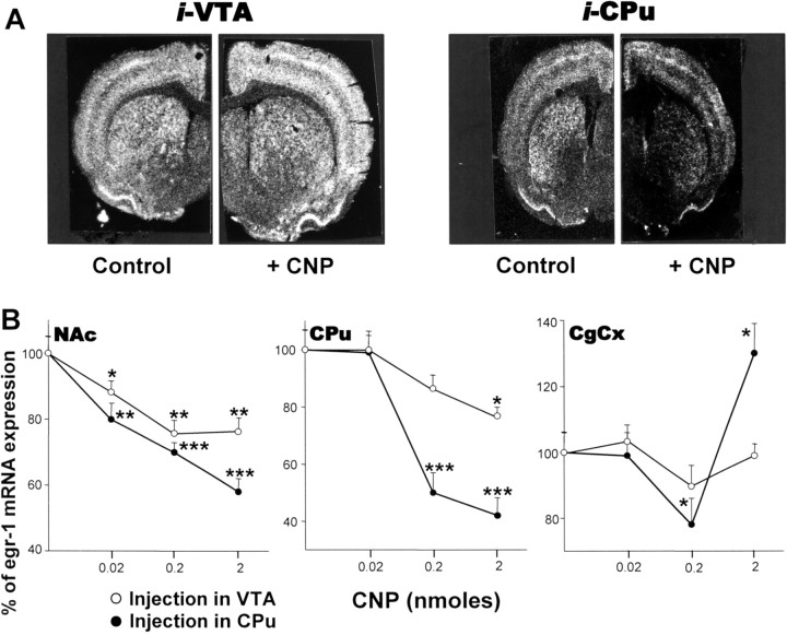
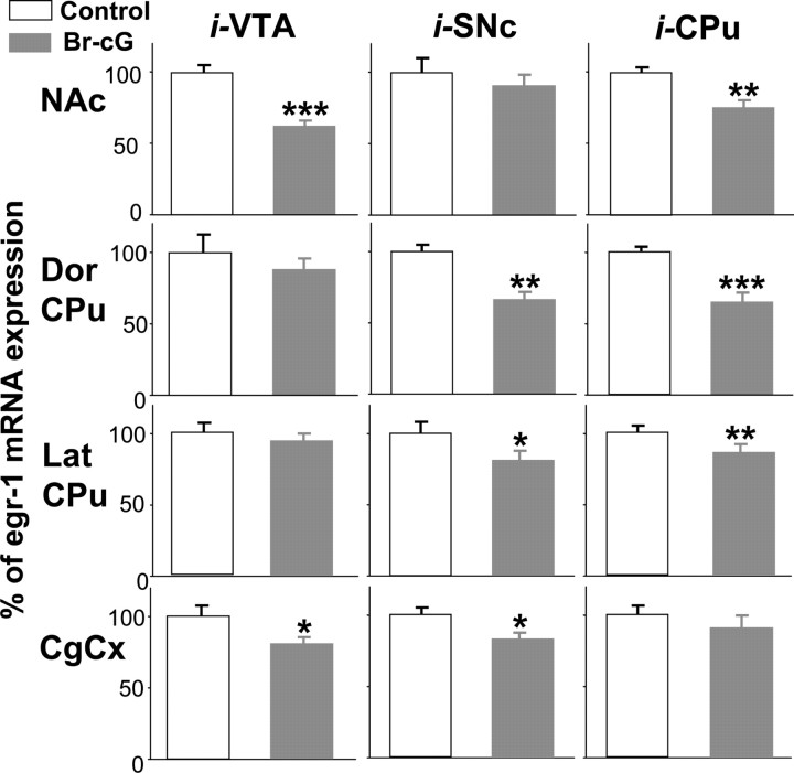
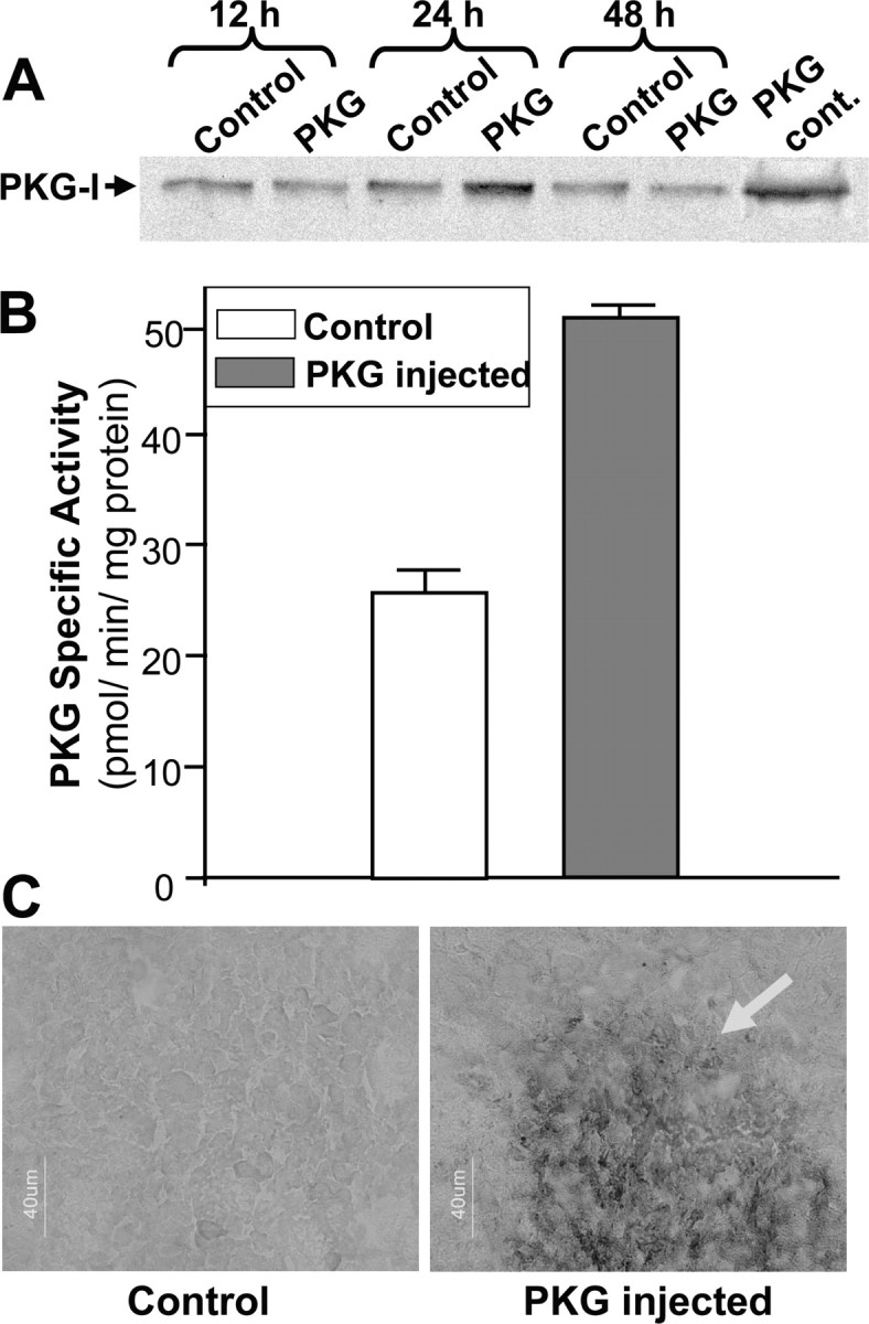
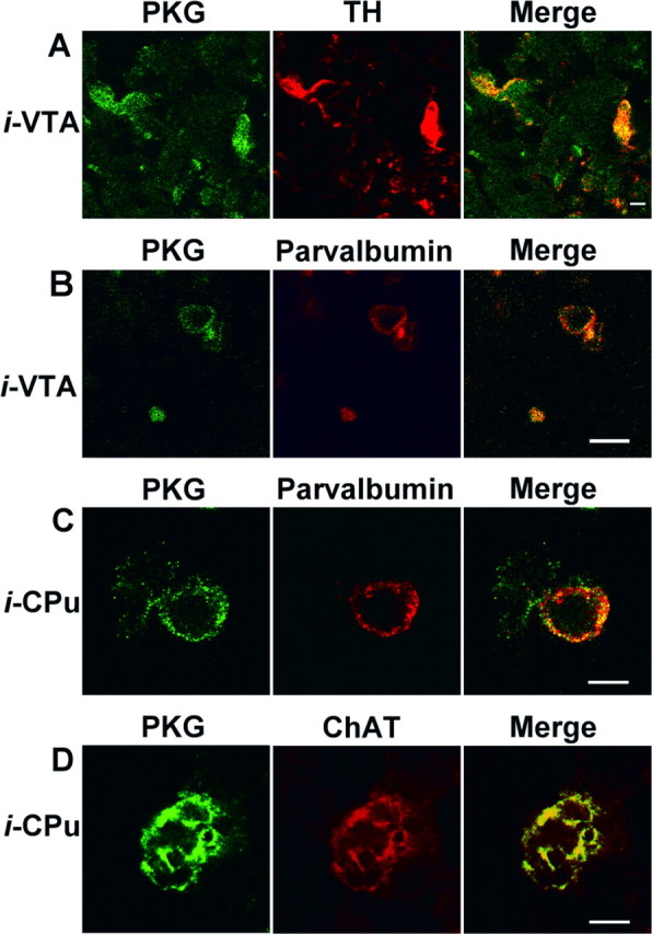
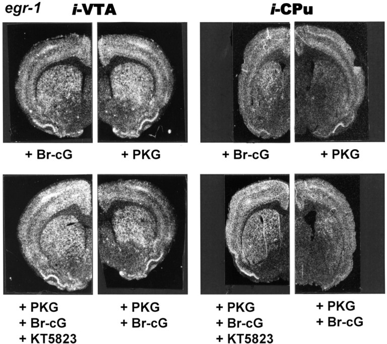
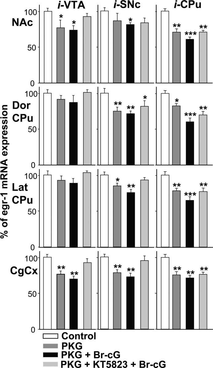
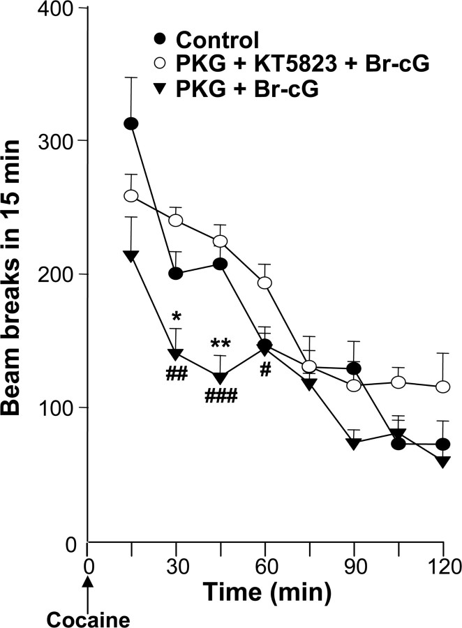
Nitric oxide (NO) and the C-type natriuretic peptide (CNP) exert their action on brain via the cGMP signaling pathway. NO, by activating soluble guanylyl cyclase, and CNP, by stimulating membrane-bound guanylyl cyclase, cause intracellular increases of cGMP, activating cGMP-dependent protein kinases (PKGs). We show here that injection of CNP into the rat ventral tegmental area strongly reduced cocaine-induced egr-1 expression in the nucleus accumbens in a dose-dependent manner. The effect of CNP was reversed by the previous injection of a selective PKG inhibitor, KT5823. Activation of PKG by 8-bromo-cGMP reduced, like CNP, cocaine-induced gene transcription in dopaminergic structures. To confirm the involvement of PKG, this was overexpressed in either the mesencephalon or the caudate-putamen. Using the polyethyleneimine delivery system, an active protein was expressed by injecting a plasmid vector containing the human PKG-Ialpha cDNA. PKG was overexpressed in dopaminergic and GABAergic neurons when the plasmid was injected in the ventral tegmental area, whereas overexpression was observed in medium spiny GABAergic neurons and in both cholinergic and GABAergic interneurons when the PKG vector was injected into the caudate-putamen. Activation of the overexpressed PKG reduced cocaine-induced egr-1 expression in dopaminergic structures and affected behavior (i.e., locomotor activity). These effects were again reversed by previous injection of the selective PKG inhibitor. The current data suggest that NO and the neuropeptide CNP are potential regulators of cocaine-related effects on behavior.


Figures

Figure 1.
Figure 1.
Schematic representation of injection schedule used for PKG overexpression. Rats were given injections of p513 vector containing the PKG-I cDNA (0.5 μg) into the right VTA (0.6 μl), SNc (0.6 μl), or CPu (2 μl) and of 0.5 μg of vector lacking a cDNA insert into the left corresponding structure. They were then given injections of 2 nmol of KT5823 or saline and 10 min later injections of 20 nmol of Br-cG or saline into the same structures. The injection volumes were 0.5 μl for VTA and SNc and 1 μl for the CPu. Finally, animals received an intraperitoneal injection of cocaine (20 mg/kg). Rats were killed, and the brains were removed 45 min after cocaine injection.
Figure 2.
Figure 2.
Effect of various doses of CNP on cocaine-induced egr-1 gene expression. A, Negative prints of in situ hybridization autoradiograms showing mRNA expression of egr-1 in rat hemibrain coronal sections (10 μm) taken at the level of the posterior striatum (∼0.3 mm anterior to bregma) and probed with [35S] antisense riboprobe. Rats were given microinjections of 2 nmol of CNP into the right VTA or CPu and microinjection of vehicle into the left corresponding structure (Control). Fifteen minutes later, rats were given intraperitoneal injections of cocaine (20 mg/kg) and killed 45 min later. B, Comparable areas of the Nac shell, the dorsal region of the Cpu, and the anterior CgCx were quantitated from in situ autoradiograms of sections from rats given injections of CNP (0.02, 0.2, or 2 nmol) into the right VTA (0.5 μl) or CPu (2 μl) or injection of vehicle into the left corresponding structure (Control). Results are expressed as a percentage of control, as means ± SEM (n = 12 sections from 3 animals). *p < 0.05, **p < 0.01, and ***p < 0.001 indicate the degree of significance when comparing CNP treatment versus the control group (ANOVA, followed by Student-Newman-Keuls multiple comparisons test).
Figure 3.
Figure 3.
Effect of Br-cG injection on cocaine-induced egr-1 gene expression. Densitometric analysis of the effect of Br-cG injection on cocaine-induced egr-1 gene expression. Comparable areas of the Nac shell, the dorsal (Dor CPu) or lateral (Lat CPu) region of the striatum, and the anterior CgCx were quantified from in situ autoradiograms of sections from rats given injections of 20 nmol of Br-cG into the right VTA (0.5 μl), SNc (0.5 μl), or CPu (2 μl) or injection of vehicle into the left corresponding structure (Control). Fifteen minutes later, rats were given intraperitoneal injections of cocaine (20 mg/kg) and killed after 45 min. Results are expressed as a percentage of control, as means ± SEM (n = 12 sections from 3 animals). *p < 0.05, **p < 0.01, and ***p < 0.001 indicate significance when comparing Br-cG treatment versus the corresponding control group (ANOVA, followed by Student-Newman-Keuls multiple comparisons test).
Figure 4.
Figure 4.
Characterization of overexpressed PKG in rat striatum. Plasmid p513 (0.5 μg, 2 μl) containing the PKG-Iα cDNA (PKG) or empty vector (Control) was microinjected into the right and left CPu, respectively. A, Western blot analysis using polyclonal anti-PKG-I antibody showing PKG-I expression as a 75,000 protein, in striata prepared at various times after plasmid injection. PKG cont., Reactivity of pure PKG from a commercial source. B, PKG-specific activity measured in the hemistriata 24 hr after plasmid injection. C, Immunohistochemical detection of PKG in the CPu 24 hr after plasmid injection. Sections were incubated in anti-PKG antibody, then with a secondary biotinylated antibody, and PKG was revealed with streptavidin-peroxidase complex, followed by staining with diaminobenzidine and H2O2. The arrow indicates the PKG overexpression sphere. Scale bar, 40 μm.
Figure 5.
Figure 5.
Double labeling for PKG and neuronal markers after PKG plasmid injection. Immunofluorescence confocal micrographs of brain cells transfected with the p513 vector containing PKG-Iα cDNA. Only cells inside the overexpression sphere were considered. A, Plasmid was injected into the VTA; brain sections were incubated with antibodies against PKG and against tyrosine hydroxylase (TH). Secondary labeled antibodies against PKG (Alexa Fluor 488; green) and TH (Cy3; red) were used. B, Plasmid was injected into the VTA; sections were incubated with antibodies against PKG and against parvalbumin. The secondary labeled antibody against PKG was as above. An antibody labeled with Alexa Fluor 568 (red) against parvalbumin was used. C, Plasmid was injected into the CPu; sections were incubated with antibodies against PKG and against parvalbumin. The secondary labeled antibodies were as in B. D, Plasmid was injected into the CPu; sections were incubated with antibodies against PKG and choline acetyltransferase (ChAT). The secondary antibodies were as in B. Scale bars: A, B, 10 μm; C, D, 5 μm.
Figure 6.
Figure 6.
Egr-1 gene expression in response to cocaine and PKG activator or inhibitor. Negative prints of in situ hybridization autoradiograms showing mRNA expression of egr-1 in rat hemibrain coronal sections taken as described in the legend to Figure 2A. Rats were given microinjections of the various compounds into the VTA or CPu with 0.5 μg of plasmid PKG, 20 nmol of Br-cG, and 2 nmol of KT5823, according to the injection schedule shown in Figure 1. They were given intraperitoneal injections of 20 mg/kg cocaine and killed 45 min after cocaine injection. The corresponding control is shown in Figure 2A.
Figure 7.
Figure 7.
Densitometric analysis of the effect of PKG overexpression on cocaine-induced egr-1 mRNA expression. Comparable areas of the Nac shell, the dorsal (Dor CPu) or lateral (Lat CPu) region of the striatum, and the anterior CgCx were quantitated from in situ autoradiograms of sections from rats given injections of 0.5 μg of plasmid p513 containing the PKG-Iα cDNA (PKG), then with 20 nmol of Br-cG and 2 nmol of KT5823 into the right VTA (0.5 μl), SNc (2 μl) or CPu (2 μl), as indicated, according to the injection protocol shown in Figure 1. Rats were given injections of the plasmid lacking an insert and injection of the various vehicle solutions into the left corresponding structure (Control). They were killed 45 min after cocaine injection. Results are expressed as a percentage of control, as means ± SEM (n = 12 sections from 3 animals). *p < 0.05, **p < 0.01, and ***p < 0.001 indicate significance levels when comparing treated versus the control group (ANOVA, followed by Student-Newman-Keuls multiple comparisons test).
Figure 8.
Figure 8.
Effect of PKG overexpression on cocaine-induced locomotor activity. Three groups of rats were given bilateral microinjections in the CPu with PKG-I cDNA plasmid (0.5 μg, 2 μl) and 20 nmol of Br-cG (PKG + Br-cG group); and 0.5 μg of plasmid, 2 nmol of KT5823, and 20 nmol of Br-cG (PKG + KT5823 + Br-cG group); or empty plasmid and saline (Control). Each group of animals received an injection of cocaine (20 mg/kg), according to the protocol shown in Figure 1. Each point represents the mean ± SEM (n = 5) of beam breaks. * or #p < 0.05; ##p < 0.01 and *** or ###p < 0.001. * indicates significance when comparing treated versus the control group; # indicates significance when comparing PKG plus KT5823 plus Br-cG versus other treatments (ANOVA, followed by Student-Newman-Keuls multiple comparisons test).

Similar articles

Cited by

References

    1. Andersson M, Konradi C, Cenci MA (2001) cAMP response element-binding protein is required for dopamine-dependent gene expression in the intact but not the dopamine-denervated striatum. J Neurosci 21: 9930-9943. - PMC - PubMed
    1. Ariano MA (1983) Distribution of components of the guanosine 3′,5′-phosphate system in rat caudate-putamen. Neuroscience 10: 707-723. - PubMed
    1. Babinski K, Haddad P, Vallerand D, McNicoll N, de Lean A, Ong H (1995) Natriuretic peptides inhibit nicotine-induced whole-cell currents and catecholamine secretion in bovine chromaffin cells: evidence for the involvement of the atrial natriuretic factor R2 receptors. J Neurochem 64: 1080-1087. - PubMed
    1. Bhat RV, Baraban JM (1993) Activation of transcription factor genes in striatum by cocaine: role of both serotonin and dopamine systems. J Pharmacol Exp Ther 267: 496-505. - PubMed
    1. Bonkale WL, Cowburn RF, Winblad B, Fastbom J (1997) Autoradiographic characterization of [3H]cGMP binding sites in the rat brain. Brain Res 763: 1-13. - PubMed

MeSH terms

LinkOut - more resources