. 2005 Jan;131(1):41-8.
doi: 10.1007/s00432-004-0619-y. Epub 2004 Sep 30.

Increased expression of ADAM family members in human breast cancer and breast cancer cell lines

Affiliations

Increased expression of ADAM family members in human breast cancer and breast cancer cell lines

Uwe Lendeckel et al. J Cancer Res Clin Oncol. 2005 Jan.

Abstract

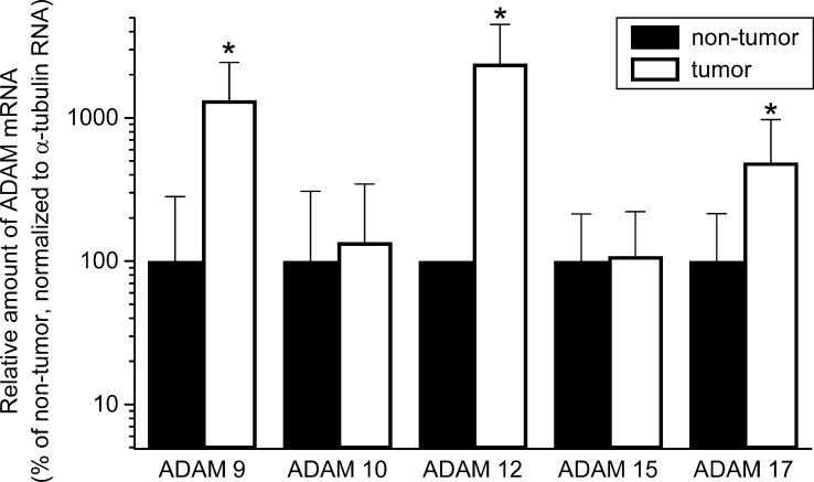
Purpose: ADAMs (A Disintegrin and Metalloprotease) are multifunctional, membrane-bound cell surface glycoproteins, which have numerous functions in cell growth, differentiation, and motility. We wished to investigate the expression of ADAM 9, 10, 12, 15, and in human breast cancer.

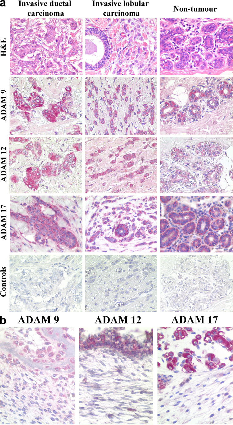
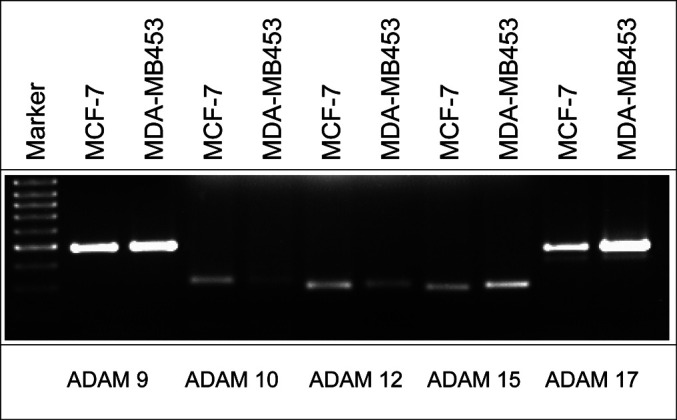
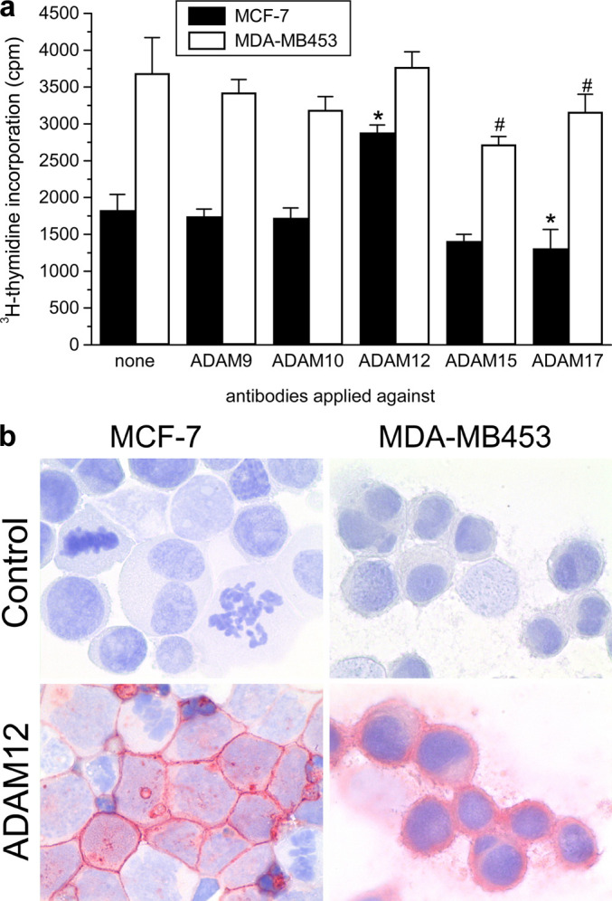
Methods: Expression of ADAMs was determined in breast cancer specimens and the corresponding non-neoplastic breast tissue from 24 patients, and in the MCF-7 and MDA-MB 453 breast cancer cell lines via quantitative RT-PCR and immunohistochemistry. The effects of anti-ADAM antibodies on cell proliferation were assessed by measuring DNA-synthesis.

Results: Breast cancer tissue samples showed increased mRNA expression of ADAM 9, 12, and 17, whereas ADAM 10 and 15 were not differently expressed. Protein expression was studied by immunohistochemistry. All ADAMs were expressed in MCF-7 and MDA-MB453 cell lines, with the highest expression levels being observed for ADAM 9, 12, and 17. Application of anti-ADAM 15 and anti-ADAM 17 antibodies significantly inhibited the proliferation of both MCF-7 and MDA-MB453 breast cancer cell lines. In contrast, the growth of MCF-7 cells appeared to be stimulated by the administration of anti-ADAM 12 antibody.

Conclusion: The results of this study suggest that ADAMs are differentially expressed in human breast cancer and are capable of modulating tumour cell growth.


Figures

Fig. 1
Fig. 1
Expression of ADAM mRNAs in human breast cancer specimens. mRNA amounts of the individual ADAMs were determined by quantitative RT-PCR in breast cancer tissue of 16 patients and compared to the corresponding non-tumorous tissue (set to 100%). Data are given as mean±SD, *P<0.05).
Fig. 2
Fig. 2
Ethidium-bromide staining of ADAM RT-PCR products separated by agarose-gel electrophoresis. Single bands of the expected size could be detected in breast cancer samples (T) or adjacent non-tumorous tissue (N).
Fig. 3
Fig. 3
Immunohistochemical Expression of ADAMs in Breast Cancer Tissue (a) and fetal tissues (b). (a) The distribution and expression pattern of ADAMs in breast carcinomas was investigated by immunohistochemistry. ADAM 9, 12, and 17 were found each in all invasive ductal and lobular carcinomas studied. The ADAMs were detected most commonly in the cytoplasm and less commonly at the cell membrane. Ductal and lobular epithelium of the non-tumorous breast also expressed ADAM 9, 12 and 17 (arrow heads; right lane). No immunostaining was found after omission of the primary antibodies (Controls). Hematoxylin and eosin (H&E). Anti-ADAM 9 (ADAM 9), anti-ADAM 12 (ADAM 12), and anti-ADAM 17 (ADAM 17). Hematoxylin counterstain; original magnifications: ×400. (b) Fetal tissue served as positive control for immunostaining with anti-ADAM 9, ADAM 12, and ADAM 17. Note expression of ADAM 9 in osteoblasts, ADAM 12 in tracheal epithelium, and of ADAM 17 in muscle cells. Hematoxylin and eosin (H&E). Anti-ADAM 9 (ADAM 9), anti-ADAM 12 (ADAM 12), and anti-ADAM 17 (ADAM 17). Hematoxylin counterstain; original magnifications: ×400.
Fig. 4
Fig. 4
Detection of ADAM mRNA expression in the human breast cancer cell lines MCF-7 and MDA-MB453. PCR products were separated by agarose-gel electrophoresis and visualised via ethidium-bromide staining
Fig. 5
Fig. 5
Effect of anti-ADAM antibodies on the growth of the human breast cancer cell lines MCF-7 and MDA-MB453 (a). Data are given as mean±SD of six experiments, *P<0.01, #P<0.05. b MCF-7 cytospins and MDA-MB435 chamber-slides were fixed and stained for ADAM12. MDA-MB435 cells exhibited a lower intensity staining for ADAM 12 with mainly cytoplasmic localisation. MCF-7 cells showed predominant surface expression of ADAM 12.

Similar articles

Cited by

References

    1. Ansorge S, Reinhold D, Lendeckel U (2003) Propolis and some of its constituents down-regulate DNA synthesis and inflammatory cytokine production but induce TGF-beta1 production of human immune cells. Z Naturforsch 58:580–589 - PubMed
    1. Asai M, Hattori C, Szabo B, Sasagawa N, Maruyama K, Tanuma S, Ishiura S (2003) Putative function of ADAM9, ADAM10, and ADAM17 as APP alpha-secretase. Biochem Biophys Res Commun 301:231–235 - PubMed
    1. Black RA and White J M (1998) ADAMs: focus on the protease domain. Curr Opin Cell Biol 10:654–659 - PubMed
    1. Black RA, Rauch CT, Kozlosky CJ, Peschon JJ, Slack JL, Wolfson MF, Castner BJ, Stocking KL, Reddy P, Srinivasan S, Nelson N, Boiani N, Schooley KA, Gerhart M, Davis R, Fitzner JN, Johnson RS, Paxton RJ, March CJ, Cerretti DP (1997) A metalloproteinase disintegrin that releases tumour-necrosis factor-alpha from cells. Nature 385:729–733 - PubMed
    1. Borrell-Pages M, Rojo F, Albanell J, Baselga J, Arribas J (2003) TACE is required for the activation of the EGFR by TGF-alpha in tumors. EMBO J 22:1114–1124 - PMC - PubMed

Publication types

MeSH terms