. 2005 Apr 15;387(Pt 2):507-17.
doi: 10.1042/BJ20041501.

MSK1 activity is controlled by multiple phosphorylation sites

Affiliations

MSK1 activity is controlled by multiple phosphorylation sites

Claire E McCoy et al. Biochem J. .

Abstract

MSK1 (mitogen- and stress-activated protein kinase) is a kinase activated in cells downstream of both the ERK1/2 (extracellular-signal-regulated kinase) and p38 MAPK (mitogen-activated protein kinase) cascades. In the present study, we show that, in addition to being phosphorylated on Thr-581 and Ser-360 by ERK1/2 or p38, MSK1 can autophosphorylate on at least six sites: Ser-212, Ser-376, Ser-381, Ser-750, Ser-752 and Ser-758. Of these sites, the N-terminal T-loop residue Ser-212 and the 'hydrophobic motif' Ser-376 are phosphorylated by the C-terminal kinase domain of MSK1, and their phosphorylation is essential for the catalytic activity of the N-terminal kinase domain of MSK1 and therefore for the phosphorylation of MSK1 substrates in vitro. Ser-381 is also phosphorylated by the C-terminal kinase domain, and mutation of Ser-381 decreases MSK1 activity, probably through the inhibition of Ser-376 phosphorylation. Ser-750, Ser-752 and Ser-758 are phosphorylated by the N-terminal kinase domain; however, their function is not known. The activation of MSK1 in cells therefore requires the activation of the ERK1/2 or p38 MAPK cascades and does not appear to require additional signalling inputs. This is in contrast with the closely related RSK (p90 ribosomal S6 kinase) proteins, whose activity requires phosphorylation by PDK1 (3-phosphoinositide-dependent protein kinase 1) in addition to phosphorylation by ERK1/2.


Figures
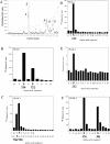
Figure 1
Figure 1. Autophosphorylation of MSK1 in vitro
Recombinant MSK1, which had been previously phosphorylated with ERK2, was autophosphorylated in vitro as described in the Methods section. The autophosphorylated MSK1 was then run on an SDS/polyacrylamide gel and the MSK1 band excised, digested with trypsin, and the resulting peptides were run on reversed-phase HPLC as described in the Methods section. The 32P elution trace is shown in (A). Phosphopeptides eluted in the 32P-labelled fractions were identified by MS, and the positions of phosphate in the peptides were determined by solid-phase sequencing for each of the peaks (BF).
Figure 2
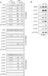
Figure 2. Phosphorylation of MSK1 in HEK-293 cells
(A) Wild-type (WT), C-terminal kinase dead (CT-KD) or N-terminal kinase dead (NT-KD) GST-tagged MSK1 was expressed in HEK-293 cells by transient transfection. Cells were serum-starved for 16 h and incubated with PD184352 (2 μM) or SB203580 (5 μM) where indicated. Cells were then stimulated with UV-C (200 J/m2, followed by 30 min at 37 °C) or PMA (TPA; 400 ng/ml, 10 min) as indicated and lysed as described in the Methods section. Lysates were immunoblotted using antibodies against GST, or MSK1 phosphorylated on Ser-212, Ser-360, Ser-376, Ser-381, Thr-581, Ser-750 or dual phosphorylated Ser-750/Ser-752. (B) Endogenous MSK1 was immunoprecipitated (IP) from HEK-293 cells and treated as indicated. Immunoprecipitates were run on SDS gels and blotted for total MSK1, phospho-Ser-212, Ser-360, Ser-376, Ser-381, Thr-581, Ser-750 and dual phosphorylated Ser-750/Ser-752.
Figure 3
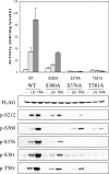
Figure 3. Ser-376 and Thr-581 phosphorylation is required for MSK1 activity
Wild-type, Ser360→Ala, Ser376→Ala or Thr581→Ala FLAG-tagged MSK1 was expressed in HEK-293 cells by transient transfection. Cells were serum-starved for 16 h and then stimulated with UV-C (200 J/m2, followed by 30 min at 37 °C) or PMA (TPA; 400 ng/ml, 10 min) as indicated and lysed as described in the Methods section. MSK1 was immunoprecipitated using an anti-FLAG antibody and assayed against a peptide substrate as described in the Methods section. The kinase activity from unstimulated cells is shown by open bars, UV-C stimulation as hatched bars and PMA stimulation as grey bars. One unit (U) of activity is defined as the amount of kinase required to incorporate 1 nmol of phosphate in 1 min. Error bars represent the S.D. for three assays. The same lysates were also immunoblotted with antibodies against FLAG or MSK1 phosphorylated on Ser-212, Ser-360, Ser-376, Ser-381 or Thr-581.
Figure 4
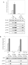
Figure 4. MSK1 autophosphorylates on Ser-212 in HEK-293 cells
(A) Wild-type MSK1 or Ser212→Ala FLAG-tagged MSK1 was expressed in HEK-293 cells by transient transfection. Cells were serum-starved for 16 h and then stimulated with UV-C (200 J/m2, followed by 30 min at 37 °C) or with PMA (400 ng/ml, 10 min) as indicated and lysed as described in the Methods section. MSK1 was immunoprecipitated using an anti-FLAG antibody and assayed against a peptide substrate as described in the Methods section. One unit (U) of activity is defined as the amount of kinase required to incorporate 1 nmol of phosphate in 1 min. The kinase activity from unstimulated cells is shown by open bars, UV-C stimulation as hatched bars and PMA stimulation as grey bars. Error bars represent the S.D. for three assays. The same lysates were also immunoblotted with antibodies against FLAG or MSK1 phosphorylated on Ser-212, Ser-360, Ser-376, Ser-381 or Thr-581. (B) Same as (A) except that GST wild-type (WT), C-terminal kinase dead (CT-KD), Ser376→Asp or C-terminal kinase dead/Ser376→Asp were expressed in HEK-293 cells. MSK1 was pulled down from 0.2 mg of lysate with glutathione–Sepharose and assayed against a peptide substrate as described in the Methods section. The same lysates were also immunoblotted with antibodies against FLAG or MSK1 phosphorylated on either Ser-212 or Ser-381.
Figure 5
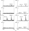
Figure 5. Phosphorylation of Ser-212 of MSK1 in HEK-293 cells
MS of the tryptic phosphopeptide (210–226 of MSK1) containing Ser-212. MSK1 [GST wild-type (A, E), C-terminal kinase dead (B, F), Ser376→Asp (C, G) or C-terminal kinase dead/Ser376→Asp (D, H)] was expressed in HEK-293 cells (control samples, AD respectively) and from a similar set of PMA-stimulated cells (+PMA samples, EH respectively). MSK1 was pulled down from 15 mg of lysate with glutathione–Sepharose and run on a 4–12% acrylamide/SDS gel. Bands corresponding to MSK1 were excised, reduced, alkylated with iodoacetamide and then digested or not with trypsin [37]. The resultant tryptic phosphopeptides were subjected to immobilized metal affinity chromatography as described in the Methods section, followed by MS on an Applied Biosystems 4700 MALDI–TOF-TOF with 0.25 μl of α-cyanocinnamic acid+10 mM NH4PO3 as the matrix. The theoretical monoisotopic mass for the peptide 210–226 (AYSFCGTIEYMAPDIVR) with one phosphoserine is 2072.89 Da, and with one phosphoserine and one oxidized methionine residue is 2088.88 Da (the cysteine residue having been converted into carbamidomethylcysteine).
Figure 6
Figure 6. Phosphorylation of Ser-381 is not essential for activity
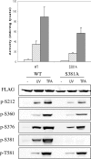
Wild-type or Ser381→Ala FLAG-tagged MSK1 was expressed in HEK-293 cells by transient transfection. Cells were serum-starved for 16 h and then stimulated with UV-C (200 J/m2, followed by 30 min at 37 °C) or PMA (400 ng/ml, 10 min) as indicated and lysed as described in the Methods section. MSK1 was immunoprecipitated using an anti-FLAG antibody and assayed against a peptide substrate as described in the Methods section. One unit (U) of activity was defined as the amount of kinase required to incorporate 1 nmol of phosphate in 1 min. The kinase activity from unstimulated cells is shown by open bars, UV-C stimulation as hatched bars and PMA stimulation as grey bars. Error bars represent the S.D. for three assays. The same lysates were also immunoblotted with antibodies against FLAG, phospho-Ser-360 MSK1, phospho-Ser-376 MSK1 or phospho-Thr-581 MSK1.
Figure 7
Figure 7. MSK1 requires a MAPK docking site for activation
(A) Wild-type, Leu740→Ala or Arg743→Ala/Arg744→Ala FLAG-tagged MSK1 was expressed in HEK-293 cells by transient transfection. Cells were serum-starved for 16 h and then stimulated with UV-C (200 J/m2, followed by 30 min at 37 °C) or PMA (400 ng/ml, 10 min) as indicated and lysed as described in the Methods section. MSK1 was immunoprecipitated using an anti-FLAG antibody and assayed against a peptide substrate as described in the Methods section. One unit (U) of activity was defined as the amount of kinase required to incorporate 1 nmol of phosphate in 1 min. The kinase activity from unstimulated cells is shown by open bars, UV-C stimulation as hatched bars and PMA stimulation as grey bars. Error bars represent the S.D. for three data points. The same lysates were also immunoblotted with antibodies against FLAG, or MSK1 phosphorylated on Ser-212, Ser-360, Ser-376, Ser-381, Thr-581, Ser-750 or dual phosphorylated Ser-750/Ser-752. (B) Same as (A) except that wild-type, Ser750→Ala/Ser752→Ala or Ser758→Ala was expressed in HEK-293 cells.
Figure 8
Figure 8. C-terminal phosphorylation of MSK1 does not affect its interaction with MAPKs
FLAG-MSK1 (wild-type, Leu740→Ala, Arg742→Ala/Arg43→Ala, Ser750→Ala/Ser752→Ala or Ser758→Ala) was transfected into HEK-293 cells. Cells were serum-starved for 16 h and then stimulated with UV-C (200 J/m2, followed by 30 min at 37 °C) or PMA (400 ng/ml, 10 min) as indicated and lysed as described in the Methods section. MSK1 was immunoprecipitated using an anti-FLAG antibody and immunoprecipitates were run on SDS gels. Gels were immunoblotted for FLAG, ERK1, ERK2 or p38α.
Figure 9
Figure 9. Sequence alignment of MSK and RSK around phosphorylation sites
The sequences around the T-loop, linker and C-terminal phosphorylation sites of MSK were aligned with RSK. Phosphorylation sites are highlighted in black and conserved residues are highlighted in grey. The hydrophobic motif in RSK is underlined and the MAPK docking site is indicated by asterisks. Black ‘lollipops’ are phosphorylation sites that are conserved between MSK and RSK isoforms. White ‘lollipops’ are phosphorylation sites that are unique to MSK1.

References

    1. Pierrat B., Correia J. d. S., Mary J.-L., Tomas-Zuber M., Lesslauer W. RSK-B, a novel ribosomal S6 kinase family member, is a CREB kinase under dominant control of p38alpha mitogen-activated protein kinase (p38alpha MAPK) J. Biol. Chem. 1998;273:29661–29671. - PubMed
    1. New L., Zhao M., Li Y., Bassett W. W., Feng Y., Ludwig S., Padova F. D., Gram H., Han J. Cloning and characterization of RLPK, a novel RSK-related protein kinase. J. Biol. Chem. 1999;274:1026–1032. - PubMed
    1. Deak M., Clifton A. D., Lucocq J. M., Alessi D. R. Mitogen- and stress-activated protein kinase-1 (MSK1) is directly activated by MAPK and SAPK2/p38, and may mediate activation of CREB. EMBO J. 1998;17:4426–4441. - PMC - PubMed
    1. Arthur J. S. C., Cohen P. MSK1 is required for CREB phosphorylation in response to mitogens in mouse embryonic stem cells. FEBS lett. 2000;482:44–48. - PubMed
    1. Wiggin G. R., Soloaga A., Foster J. M., Murray-Tait V., Cohen P., Arthur J. S. MSK1 and MSK2 are required for the mitogen- and stress-induced phosphorylation of CREB and ATF1 in fibroblasts. Mol. Cell. Biol. 2002;22:2871–2881. - PMC - PubMed

Publication types

MeSH terms

Substances