

Low nNOS protein in the locus coeruleus in major depression
- PMID: 15569249
- PMCID: PMC2923201
- DOI: 10.1111/j.1471-4159.2004.02792.x
Low nNOS protein in the locus coeruleus in major depression
Abstract
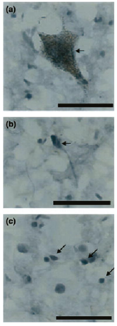
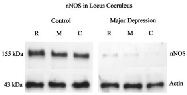
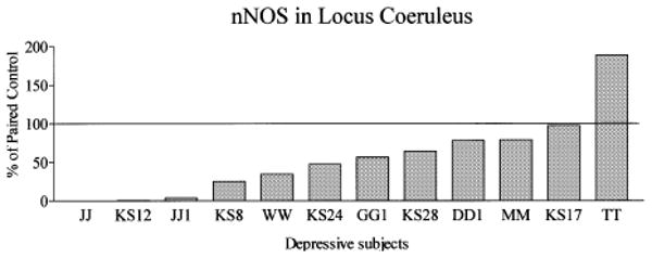
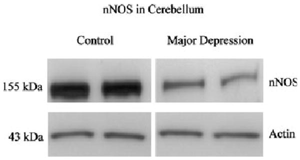
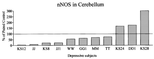
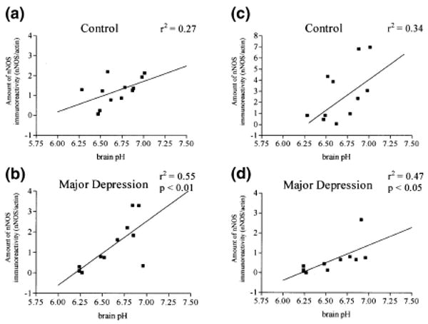
Disruptions of glutamatergic and noradrenergic signaling have been postulated to occur in depressive disorders. Glutamate provides excitatory input to the noradrenergic locus coeruleus (LC). In this study, the location of immunoreactivity against neuronal nitric oxide synthase (nNOS), an intracellular mediator of glutamate receptor activation, was examined in the normal human LC, and potential changes in nNOS immunoreactivity that might occur in major depression were evaluated. Tissue containing LC, and a non-limbic, LC projection area (cerebellum) was obtained from 11 to 12 matched pairs of subjects with major depression and control subjects lacking major psychiatric diagnoses. In the LC region, nNOS immunoreactivity was found in large neuromelanin-containing neurons, small neurons lacking neuromelanin, and glial cells. Levels of nNOS immunoreactivity were significantly lower in the LC (- 44%, p < 0.05), but not in the cerebellum, when comparing depressed with control subjects. nNOS levels were positively correlated with brain pH values in depressed, but not control, subjects in both brain regions. Low levels of nNOS in the LC may reflect altered excitatory input to this nucleus in major depression. However, pH appears to effect preservation of nNOS immunoreactivity in subjects with depression. This factor may contribute, in part, to low levels of nNOS in depression.
Figures
References
-
- Allgaier C, Durmaz M, Muller D, Franke H, Poelchen W, Wirkner K, Illes P. Single-cell RT-PCR analysis of N-methyl-d-aspartate receptor subunit expression in rat locus coeruleus neurones. Naunyn Schmiedebergs Arch Pharmacol. 2001;363:120–123. - PubMed
-
- Altamura CA, Mauri MC, Ferrara A, Moro AR, D'Andrea G, Zamberlan F. Plasma and platelet excitatory amino acids in psychiatric disorders. Am J Psychiatry. 1993;150:1731–1733. - PubMed
-
- Altamura C, Maes M, Dai J, Meltzer HY. Plasma concentrations of excitatory amino acids, serine, glycine, taurine and histidine in major depression. Eur Neuropsychopharmacol. 1995;5:71–75. - PubMed
-
- Aoki E, Semba R, Mikoshiba K, Kashiwamata S. Predominant localization in glial cells of free 1-arginine: immunocytochemical evidence. Brain Res. 1991;547:190–192. - PubMed
-
- Arbones ML, Ribera J, Agullo L, Baltrons MA, Casanovas A, Riveros-Moreno V, Garcia A. Characteristics of nitric oxide synthase type I of rat cerebellar astrocytes. Glia. 1996;18:224–232. - PubMed
Publication types
MeSH terms
Substances
Grants and funding
LinkOut - more resources
Full Text Sources
