. 2004 Dec 6;167(5):935-44.
doi: 10.1083/jcb.200409107. Epub 2004 Nov 29.

Progenitor cells of the testosterone-producing Leydig cells revealed

Affiliations

Progenitor cells of the testosterone-producing Leydig cells revealed

Michail S Davidoff et al. J Cell Biol. .

Abstract

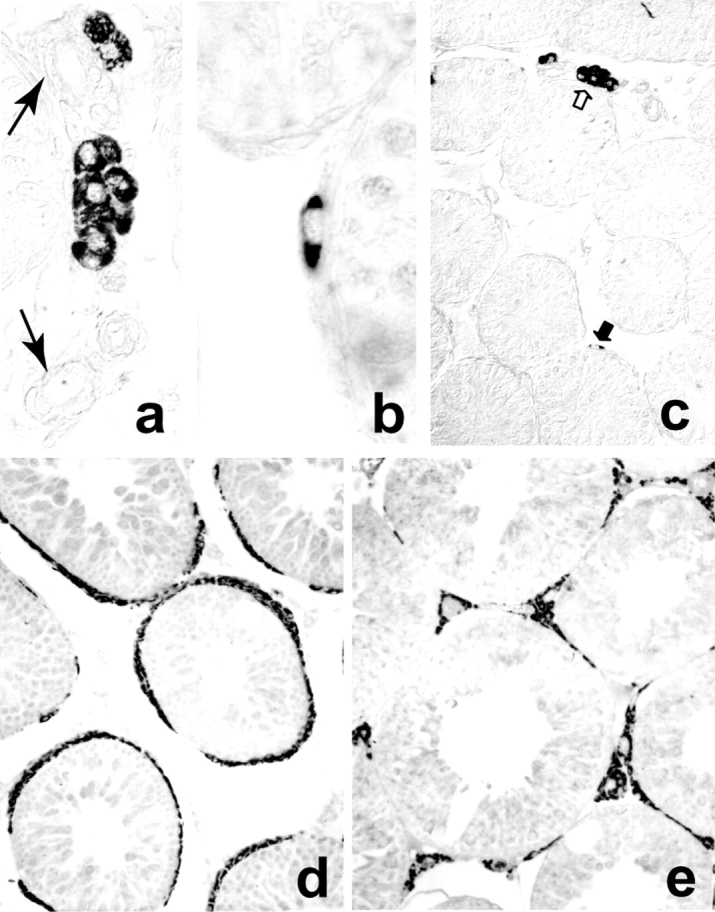
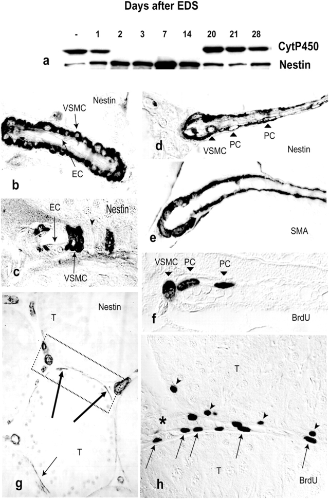
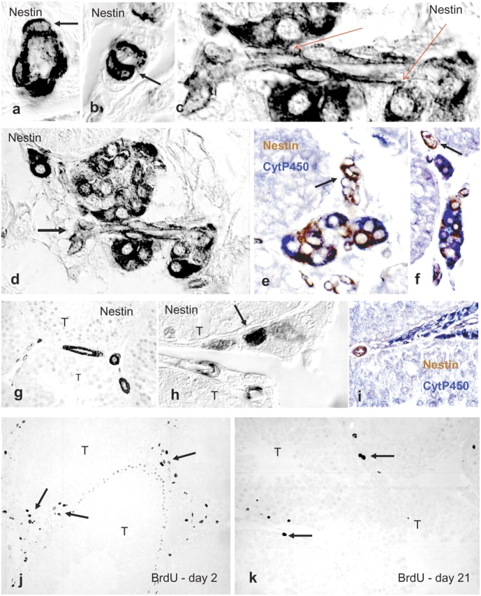
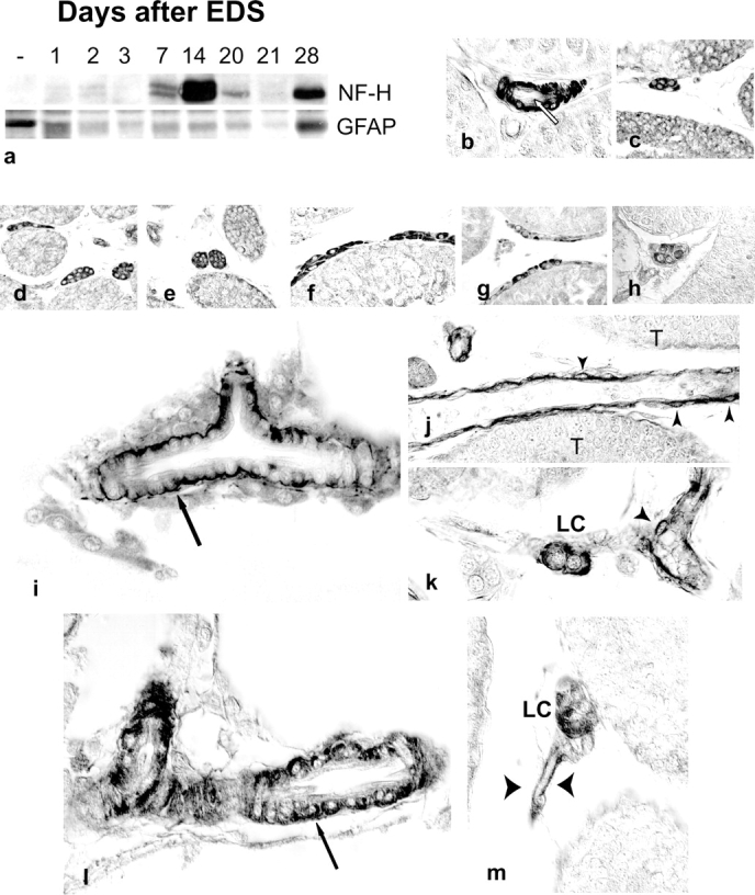
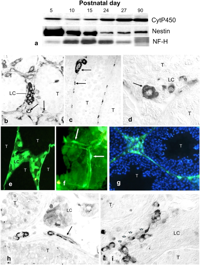
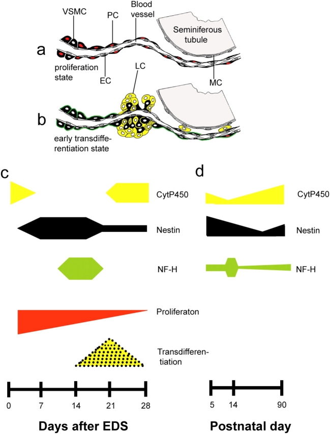
The cells responsible for production of the male sex hormone testosterone, the Leydig cells of the testis, are post-mitotic cells with neuroendocrine characteristics. Their origin during ontogeny and regeneration processes is still a matter of debate. Here, we show that cells of testicular blood vessels, namely vascular smooth muscle cells and pericytes, are the progenitors of Leydig cells. Resembling stem cells of the nervous system, the Leydig cell progenitors are characterized by the expression of nestin. Using an in vivo model to induce and monitor the synchronized generation of a completely new Leydig cell population in adult rats, we demonstrate specific proliferation of vascular progenitors and their subsequent transdifferentiation into steroidogenic Leydig cells which, in addition, rapidly acquire neuronal and glial properties. These findings, shown to be representative also for ontogenetic Leydig cell formation and for the human testis, provide further evidence that cellular components of blood vessels can act as progenitor cells for organogenesis and repair.


Figures

Figure 1.
Figure 1.
Reappearance of Leydig cells after EDS treatment. Leydig (CytP450-immunoreactive) cells are first detectable near blood vessels (arrows) in the form of clusters (a) or as single spindle-shaped cells (b) in testes 14 d after EDS. The cluster (white arrow) and spindle-shaped cell (black arrow) magnified in a and b, respectively, are indicated in c. The accumulation of Leydig cells at d 21 (d) and d 30 (e) after EDS, distributed around (d) and between (e) seminiferous tubules, is shown.
Figure 2.
Figure 2.
EDS exposure induces testicular nestin expression, localized to VSMCs and PCs. (a) Immunoblots demonstrate increased nestin expression during the period of Leydig cell (see levels of CytP450) depletion. (b–d) Immunohistochemical analyses show that nestin is localized to vascular smooth muscle cells (VSMC) and pericytes (PC) (d), but not to endothelial cells (EC; see b and c, with longitudinal orientation) of testicular blood vessels. (e) VSMCs and PCs are stained for smooth muscle α-actin (SMA). (f–h) At d 2 after EDS, VSMCs and PCs proliferate as indicated by nuclear incorporation of BrdU (f and h). The testicular distribution of nestin immunoreactivity at that time, localized to large intertubular as well as to peritubular (arrows) vessels, is shown in g. Higher magnification of an area corresponding to the box marked in g demonstrates proliferating (BrdU labeled) cells of the peritubular microvasculature (h, arrows). Note that some spermatogonia (arrowheads) are also detectable. The lumen of a blood vessel filled with erythrocytes is marked by an asterisk. T, seminiferous tubules.
Figure 3.
Figure 3.
Nestin-expressing cells protrude from blood vessel walls and transform into Leydig cells. 14 d after EDS, nestin-immunoreactive cells begin to protrude (arrows) from intertubular vessel walls (a and b) and begin to form cell clusters in their direct vicinity (c and d). The orientation of such a vessel between newly formed cell clusters (d, indicated by an arrow) and sites where transdifferentiating progenitor cells are still in contact with the vessel they derive from (c, red arrows) are shown. These clusters consist of cells that either coexpress nestin (brown) and CytP450 (blue), or express Cyt P450 only (e and f). 1 wk later, nestin+ cells are visible at a peritubular position (g and h), and newly formed Leydig cells are only occasionally (h, day 20) or not at all (i, day 21) nestin positive. At this time (k, day 21), the number of proliferating (BrdU labeled) vascular wall cells (arrows) is much lower than at d 2 after EDS (j). T, seminiferous tubules.
Figure 4.
Figure 4.
Neural characteristics of Leydig cells and their vascular progenitors. (a) Immunoblots show transiently elevated levels of NF-H and a reduced expression of GFAP in testis during the period of Leydig cell depletion after EDS treatment. VSMCs, but not endothelial cells (EC, marked by an arrow), are the sites of NF-H expression (b). In contrast, newly formed Leydig cells are immunoreactive for GFAP (c), GAP43 (d), TrkA (e and f), GDNF (g), and N-CAM (h). Both VSMCs (i, arrow) and PCs (j and k, arrowheads), as well as newly formed Leydig cells (k, LC), are characterized by the expression of NG2, and Leydig cells (LC) together with their vascular progenitors (VSMC, arrow; PCs, arrowheads) are sites of PDGFR-β expression (l and m). T, seminiferous tubules.
Figure 5.
Figure 5.
Nestin expression in testis during rat ontogenetic development, in nestin-GFP transgenic mice, and adult humans. (a) Immunoblots show the regulation in rat testis of CytP450, nestin, and NF-H during postnatal development. (b–d) The distribution of nestin-immunoreactive structures in rat testis at postnatal days 5 (b), 27 (c) and 90 (d) is shown. (e–g) GFP expression in testes of transgenic mice expressing GFP under the control of the nestin second-intron enhancer (green fluorescence) is detectable in Leydig cells and their vascular progenitors based on morphology (arrows point to PCs). In g, nuclei are additionally marked by DAPI (blue). (h and i) Immunohistochemical staining for nestin on adult human testis sections from individuals without (h) or with (i) Leydig cell hyperplasia. Tubular compartments (T), Leydig cells (LC), and nestin-positive vessels (arrows) are indicated. In i, stars mark the lumen of a small vessel.
Figure 6.
Figure 6.
Schematic representation of transdifferentiation of vascular progenitors into Leydig cells. (a) Nestin-expressing (black) vascular smooth muscle cells (VSMC) and pericytes (PC) of testicular blood vessels, but not endothelial cells (EC) or peritubular myoid cells (MC), vigorously proliferate (indicated by red-labeled nuclei) during the first week after EDS-induced Leydig cell depletion. (b) Around d 14 after EDS, nestin-expressing VSMCs and PCs, now marked also by a transient expression of NF-H (green contour line), begin to protrude from intertubular vessels and begin to form cell clusters. By transdifferentiation, the cells first acquire steroidogenic properties (yellow, black dotted) and finally loose nestin, resulting in typical (yellow) Leydig cells (LC). (c and d) Time scale of Leydig cell (re)generation after EDS treatment (c) and during ontogenetic development (d). (c) Leydig cell depletion, marked by the absence of CytP450 (yellow), rapidly induces nestin expression (black) and proliferation activity (red). The conversion of proliferative into transformation activity (yellow, black dotted) is preceded by a transient expression of NF-H (green). (d) A similar correlation between CytP450, nestin, and NF-H expression is detectable during postnatal testis development.

Similar articles

Cited by

References

    1. Alliot, F., J. Rutin, P.J.M. Leenen, and B. Pessac. 1999. Pericytes and periendothelial cells of brain parenchyma vessels co-express aminopeptidase N, aminopeptidase A, and nestin. J. Neurosci. Res. 58:367–378. - PubMed
    1. Allt, G., and J.G. Lawrenson. 2001. Pericytes: cell biology and pathology. Cells Tissues Organs. 169:1–11. - PubMed
    1. Amoh, Y., L. Ligna, M. Yang, A.R. Moossa, K. Katsuoka, S. Penman, and R.M. Hoffman. 2004. Nascent blood vessels in the skin arise from nestin-expressing hair-follicle cells. Proc. Natl. Acad. Sci. USA. 101:13291–13295. - PMC - PubMed
    1. Ariyaratne, H.B.S., S.M.L.C. Mendis-Handagama, D.B. Hales, and J.I. Mason. 2000. Studies on the onset of Leydig precursor cell differentiation in the prepubertal rat testis. Biol. Reprod. 63:165–171. - PubMed
    1. Basciani, S., S. Mariani, M. Arizzi, S. Ulisse, N. Rucci, E.A. Jannini, C.D. Rocca, A. Manicone, C. Carani, G. Spera, and L. Gnessi. 2002. Expression of platelet-derived growth factor-A (PDGF-A), PDGF-B, and PDGF receptor-α and -β during human testicular development and disease. J. Clin. Endocrinol. Metab. 87:2310–2319. - PubMed

Publication types

MeSH terms