The neuronal scaffold protein Shank3 mediates signaling and biological function of the receptor tyrosine kinase Ret in epithelial cells
- PMID: 15569713
- PMCID: PMC2172453
- DOI: 10.1083/jcb.200404108
The neuronal scaffold protein Shank3 mediates signaling and biological function of the receptor tyrosine kinase Ret in epithelial cells
Abstract
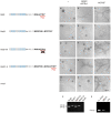
Shank proteins, initially also described as ProSAP proteins, are scaffolding adaptors that have been previously shown to integrate neurotransmitter receptors into the cortical cytoskeleton at postsynaptic densities. We show here that Shank proteins are also crucial in receptor tyrosine kinase signaling. The PDZ domain-containing Shank3 protein was found to represent a novel interaction partner of the receptor tyrosine kinase Ret, which binds specifically to a PDZ-binding motif present in the Ret9 but not in the Ret51 isoform. Furthermore, we show that Ret9 but not Ret51 induces epithelial cells to form branched tubular structures in three-dimensional cultures in a Shank3-dependent manner. Ret9 but not Ret51 has been previously shown to be required for kidney development. Shank3 protein mediates sustained Erk-MAPK and PI3K signaling, which is crucial for tubule formation, through recruitment of the adaptor protein Grb2. These results demonstrate that the Shank3 adaptor protein can mediate cellular signaling, and provide a molecular mechanism for the biological divergence between the Ret9 and Ret51 isoform.
Figures


Similar articles
-
Differential requirement of Tyr1062 multidocking site by RET isoforms to promote neural cell scattering and epithelial cell branching.Oncogene. 2004 Sep 23;23(44):7297-309. doi: 10.1038/sj.onc.1207862. Oncogene. 2004. PMID: 15326489
-
Distinct turnover of alternatively spliced isoforms of the RET kinase receptor mediated by differential recruitment of the Cbl ubiquitin ligase.J Biol Chem. 2005 Apr 8;280(14):13442-9. doi: 10.1074/jbc.M500507200. Epub 2005 Jan 27. J Biol Chem. 2005. PMID: 15677445
-
Biochemical and biological responses induced by coupling of Gab1 to phosphatidylinositol 3-kinase in RET-expressing cells.Biochem Biophys Res Commun. 2004 Oct 8;323(1):345-54. doi: 10.1016/j.bbrc.2004.08.095. Biochem Biophys Res Commun. 2004. PMID: 15351743
-
Signaling by Kit protein-tyrosine kinase--the stem cell factor receptor.Biochem Biophys Res Commun. 2005 Nov 11;337(1):1-13. doi: 10.1016/j.bbrc.2005.08.055. Biochem Biophys Res Commun. 2005. PMID: 16129412 Review.
-
Assembly of PRR-containing receptors on scaffolds: a model for imidazoline I(1)-receptor action.Ann N Y Acad Sci. 2003 Dec;1009:413-8. doi: 10.1196/annals.1304.055. Ann N Y Acad Sci. 2003. PMID: 15028620 Review.
Cited by
-
Molecular handoffs in nitrergic neurotransmission.Front Med (Lausanne). 2014 Apr 10;1:8. doi: 10.3389/fmed.2014.00008. eCollection 2014. Front Med (Lausanne). 2014. PMID: 25705621 Free PMC article. Review.
-
RET revisited: expanding the oncogenic portfolio.Nat Rev Cancer. 2014 Mar;14(3):173-86. doi: 10.1038/nrc3680. Nat Rev Cancer. 2014. PMID: 24561444 Review.
-
Transcriptional and post-translational changes in the brain of mice deficient in cholesterol removal mediated by cytochrome P450 46A1 (CYP46A1).PLoS One. 2017 Oct 26;12(10):e0187168. doi: 10.1371/journal.pone.0187168. eCollection 2017. PLoS One. 2017. PMID: 29073233 Free PMC article.
-
Organotypic specificity of key RET adaptor-docking sites in the pathogenesis of neurocristopathies and renal malformations in mice.J Clin Invest. 2010 Mar;120(3):778-90. doi: 10.1172/JCI41619. Epub 2010 Feb 15. J Clin Invest. 2010. PMID: 20160347 Free PMC article.
-
Expression of postsynaptic density proteins of the ProSAP/Shank family in the thymus.Histochem Cell Biol. 2006 Dec;126(6):679-85. doi: 10.1007/s00418-006-0199-9. Epub 2006 Jun 7. Histochem Cell Biol. 2006. PMID: 16758162
References
-
- Alberti, L., M.G. Borrello, S. Ghizzoni, F. Torriti, M.G. Rizzetti, and M.A. Pierotti. 1998. Grb2 binding to the different isoforms of Ret tyrosine kinase. Oncogene. 17:1079–1087. - PubMed
-
- Arighi, E., L. Alberti, F. Torriti, S. Ghizzoni, M.G. Rizzetti, G. Pelicci, B. Pasini, I. Bongarzone, C. Piutti, M.A. Pierotti, and M.G. Borrello. 1997. Identification of Shc docking site on Ret tyrosine kinase. Oncogene. 14:773–782. - PubMed
-
- Baloh, R.H., M.G. Tansey, E.M. Johnson Jr., and J. Milbrandt. 2000. Functional mapping of receptor specificity domains of glial cell line-derived neurotrophic factor (GDNF) family ligands and production of GFRalpha1 RET-specific agonists. J. Biol. Chem. 275:3412–3420. - PubMed
-
- Besset, V., R.P. Scott, and C.F. Ibanez. 2000. Signaling complexes and protein-protein interactions involved in the activation of the Ras and phosphatidylinositol 3-kinase pathways by the c-Ret receptor tyrosine kinase. J. Biol. Chem. 275:39159–39166. - PubMed
-
- Bockers, T.M., M.G. Mameza, M.R. Kreutz, J. Bockmann, C. Weise, F. Buck, D. Richter, E.D. Gundelfinger, and H.J. Kreienkamp. 2001. Synaptic scaffolding proteins in rat brain. Ankyrin repeats of the multidomain Shank protein family interact with the cytoskeletal protein alpha-fodrin. J. Biol. Chem. 276:40104–40112. - PubMed
MeSH terms
Substances
LinkOut - more resources
Full Text Sources
Other Literature Sources
Molecular Biology Databases
Research Materials
Miscellaneous

