. 2004 Dec 6;167(5):863-74.
doi: 10.1083/jcb.200407022. Epub 2004 Nov 29.

Phosphate deprivation induces transfer of DGDG galactolipid from chloroplast to mitochondria

Affiliations

Phosphate deprivation induces transfer of DGDG galactolipid from chloroplast to mitochondria

Juliette Jouhet et al. J Cell Biol. .

Abstract

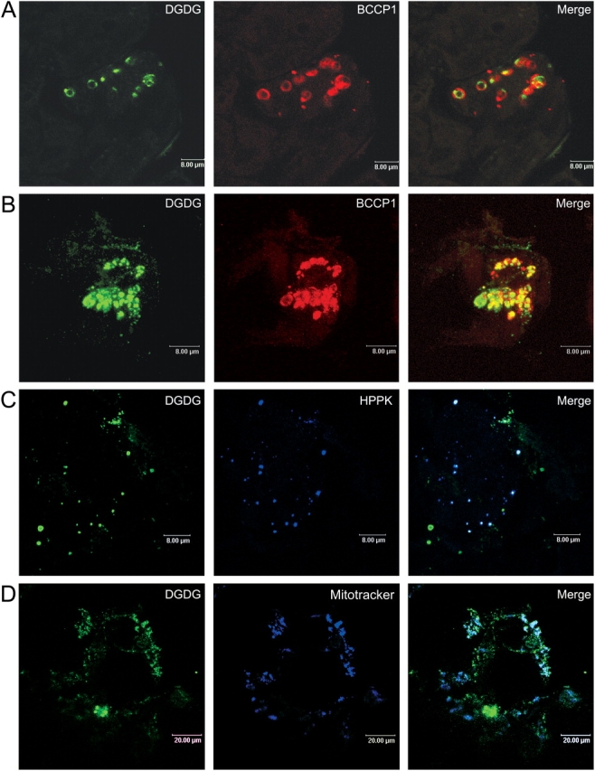
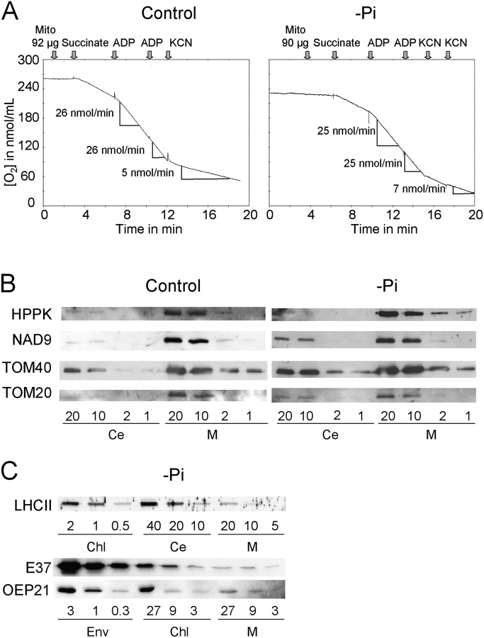
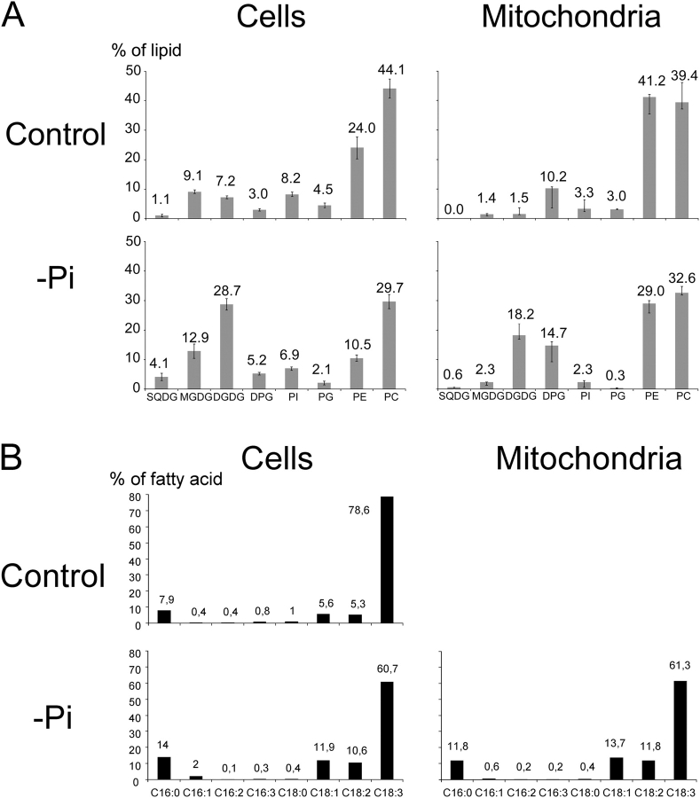
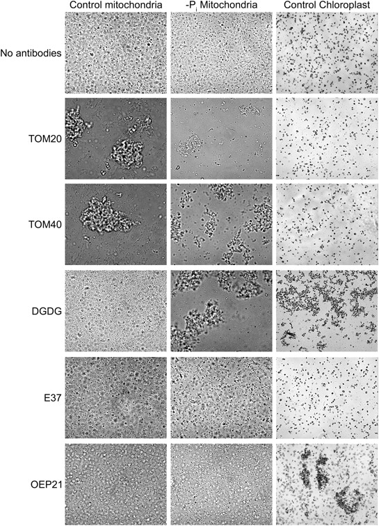
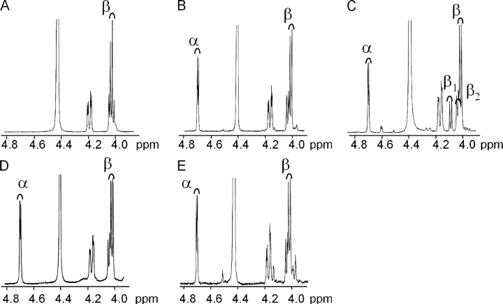
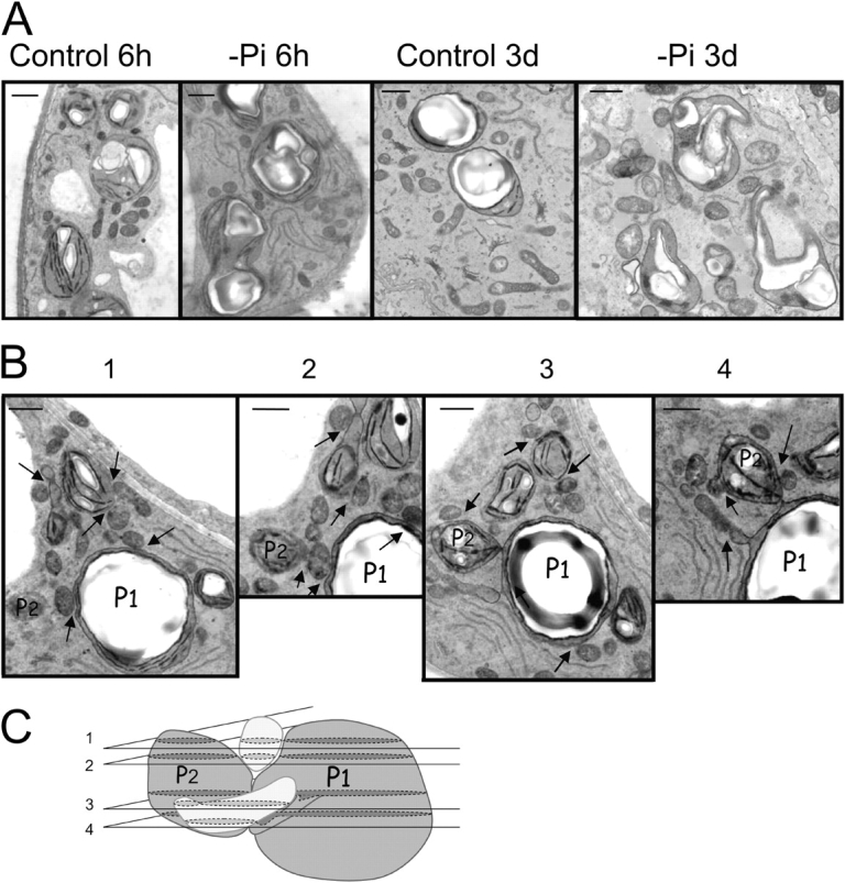
In many soils plants have to grow in a shortage of phosphate, leading to development of phosphate-saving mechanisms. At the cellular level, these mechanisms include conversion of phospholipids into glycolipids, mainly digalactosyldiacylglycerol (DGDG). The lipid changes are not restricted to plastid membranes where DGDG is synthesized and resides under normal conditions. In plant cells deprived of phosphate, mitochondria contain a high concentration of DGDG, whereas mitochondria have no glycolipids in control cells. Mitochondria do not synthesize this pool of DGDG, which structure is shown to be characteristic of a DGD type enzyme present in plastid envelope. The transfer of DGDG between plastid and mitochondria is investigated and detected between mitochondria-closely associated envelope vesicles and mitochondria. This transfer does not apparently involve the endomembrane system and would rather be dependent upon contacts between plastids and mitochondria. Contacts sites are favored at early stages of phosphate deprivation when DGDG cell content is just starting to respond to phosphate deprivation.


Figures

Figure 1.
Figure 1.
Localization of DGDG in mitochondria of A. thalia na cells deprived of Pi for 3 d. Cells (A, control; B–D, Pi-deprived) were processed for indirect immunofluorescence labeling using anti-DGDG with secondary antibodies coupled to BODIPY and either anti-BCCP1, for chloroplast detection (A and B), or anti-HPPK (C), for mitochondria detection, with secondary antibodies coupled to Alexa 594. In D, mitochondria were visualized by staining with Mitotracker orange CMTMRos. Cells were observed by confocal microscopy. Bars: A–C, 8 μm; D, 20 μm.
Figure 2.
Figure 2.
Characterization of mitochondria fractions isolated from either control or 3 d Pi-deprived A. thaliana cells. (A) To check purity and intactness of isolated mitochondria, succinate oxidation was followed by measuring O2 consumption. On average, each purified fraction consumed ∼280 nmol O2.min−1.mg−1 protein in the presence of succinate and ADP, and O2 consumption was stimulated 2.4 times by addition of ADP. Therefore, fractions were considered to be highly enriched in functionally intact mitochondria. Cyanide resistant pathway was slightly enhanced in Pi-deprived conditions as expected according to Rébeillé et al. (1984). (B) Comparative Western blot analysis of mitochondrial (M) and total cell extract (Ce) using antibodies specific for mitochondrial proteins, HPPK, a matrix protein, NAD9, an inner membrane protein, and TOM20 and TOM40 outer membrane proteins. (C) Western blot analysis of mitochondrial (M), chloroplast (Chl), and total cell extract (Ce) of Pi-deprived cells and of chloroplast envelope (Env) prepared from Arabidopsis plants as in Awai et al. (2001) using antibodies specific for chloroplast membrane proteins, LHCII for thylakoid, E37 for inner envelope membrane, and OEP21 for outer envelope membrane.
Figure 3.
Figure 3.
Glycerolipid analysis of total cell and mitochondria fractions from control and 3 d P i -deprived A. thaliana cells. (A) Glycerolipid composition. SD was calculated on four independent measurements in each case. (B) Fatty acid composition of DGDG isolated either from total cell extracts from 3 d Pi-deprived cells and control cells or from mitochondria fraction from 3 d Pi-deprived cells.
Figure 4.
Figure 4.
Immunoagglutination assays of isolated mitochondria prepared from either control or 3 d Pi-deprived A. thaliana cells. Mitochondria from Pi-deprived cells, control cells, and chloroplasts were incubated with antibodies as specified. Addition of anti-DGDG lead to agglutination of chloroplasts and of mitochondria of Pi-deprived cells only. Control antibodies were directed against E37 and OEP21 chloroplast inner and outer membrane protein, respectively, and TOM20 and TOM40 outer membrane mitochondrial proteins.
Figure 5.
Figure 5.
1H-NMR galactolipid analysis. The α-peak is characterized by a doublet at 4.7 ppm and the β-peak by a doublet at 4.0 ppm. (A) β-MGDG from A. thaliana control cells. (B) α-β DGDG from A. thaliana control cells. (C) DGDG from Spinacia oleracea purified chloroplast envelope. In envelope fraction, two DGDG types are visible: α-β and β12. (D) α-β DGDG from A. thaliana Pi-deprived cells. (E) Mitochondrial α-β DGDG from A. thaliana Pi-deprived cells.
Figure 6.
Figure 6.
Electron microscopy observation of chloroplasts and mitochondria in A. thaliana cells grown in suspension. (A) Comparison of cells grown for different times (6 h or 3 d) in standard or −Pi medium. (B) Serial cross sections of a cell grown for 6 h in −Pi medium. Arrows indicate position of contact between mitochondria and chloroplasts. (C) Three-dimensional interpretation of relative position of chloroplasts P1 and P2 (referring to P1 and P2 in B) and two mitochondria (in light gray). Numbers of cross sections refer to numbers displayed in B. Bars, 1 μm.

References

    1. Achleitner, G., B. Gaigg, A. Krasser, E. Kainersdorfer, S.D. Kohlwein, A. Perktold, G. Zellnig, and G. Daum. 1999. Association between the endoplasmic reticulum and mitochondria of yeast facilitates interorganelle transport of phospholipids through membrane contact. Eur. J. Biochem. 264:545–553. - PubMed
    1. Alban, C., P. Baldet, and R. Douce. 1994. Localization and characterization of two structurally different forms of acetyl-CoA carboxylase in young pea leaves, of which one is sensitive to aryloxyphenoxypropionate herbicides. Biochem. J. 300:557–565. - PMC - PubMed
    1. Andersson, M.X., M.H. Stridh, K.E. Larsson, C. Liljenberg, and A.S. Sandelius. 2003. Phosphate-deficient oat replaces a major portion of the plasma membrane phospholipids with the galactolipid digalactosyldiacylglycerol. FEBS Lett. 537:128–132. - PubMed
    1. Ardail, D., F. Lerme, and P. Louisot. 1991. Involvement of contact sites in phosphatidylserine import into liver mitochondria. J. Biol. Chem. 266:7978–7981. - PubMed
    1. Awai, K., E. Maréchal, M.A. Block, D. Brun, T. Masuda, H. Shimada, K. Takamiya, H. Ohta, and J. Joyard. 2001. Two types of MGDG synthase genes, found widely in both 16:3 and 18:3 plants, differentially mediate galactolipid syntheses in photosynthetic and nonphotosynthetic tissues in Arabidopsis thaliana. Proc. Natl. Acad. Sci. USA. 98:10960–10965. - PMC - PubMed

MeSH terms