

Assessment of transient ischemic attack with diffusion- and perfusion-weighted imaging
- PMID: 15569725
- PMCID: PMC8148750
Assessment of transient ischemic attack with diffusion- and perfusion-weighted imaging
Abstract
Background: Diagnosing TIA can be difficult, since evidence of brain ischemia is habitually lacking on CT and conventional MR imaging. It has been suggested that patients with acute brain infarction on neuroimaging should be considered stroke cases instead of TIA, regardless of duration of symptoms, implying that optimal diagnostic methods need to be utilized. We therefore postulated that perfusion-weighted MR imaging (PW imaging) would be useful in the diagnosis of TIA.
Methods: Retrospective analysis of 22 patients with reversible neurologic symptoms lasting less than 24 hours, assessed with DW and PW imaging.
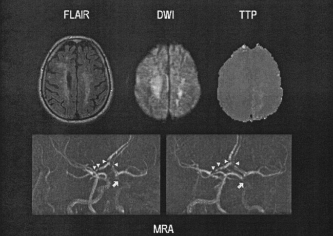
Results: MR imaging was abnormal in 15 patients (68%): 12 had abnormal DW imaging, four had both DW and PW imaging defects (all with a mismatch) and three had an isolated PW imaging abnormality. There were no differences in symptom duration, stroke etiology or cardiovascular risk factors between patients with abnormal MR imaging and those with unremarkable scan. Patients with mismatch were more likely to need conventional angiography or other cerebrovascular procedures.
Conclusion: The combined use of DW imaging and PW imaging provided evidence of brain ischemia in most patients with clinical diagnosis of TIA. Prospective studies using follow-up MR imaging are required to determine the outcome of affected tissue, as well as the clinical implications of DW-PW imaging abnormalities.
Figures


References
-
- Albers GW, Caplan LR, Easton JD, Fayad PB, Mohr JP, Saver JL, Sherman DG, the TIA Working Group. Transient ischemic attack–proposal for a new definition. N Engl J Med 2002;347:1713–1716 - PubMed
-
- Kidwell CS, Alger JR, Di Salle F, Starkman S, Villablanca P, Bentson J, Saver JL. Diffusion MRI in patients with transient ischemic attacks. Stroke 1999;30:1174–1180 - PubMed
-
- Ay H, Oliveira-Filho J, Buonanno FS, et al. ’Footprints’ of transient ischemic attacks: a diffusion-weighted MRI study. Cerebrovasc Dis 2002;14:177–186 - PubMed
-
- Crisostomo RA, Garcia MM, Tong DC. Detection of diffusion-weighted MRI abnormalities in patients with transient ischemic attack: correlation with clinical characteristics. Stroke 2003;34:932–937 - PubMed
Publication types
MeSH terms
Grants and funding
LinkOut - more resources
Full Text Sources
Medical