. 2004 Nov-Dec;25(10):1688-95.

Acute methotrexate neurotoxicity: findings on diffusion-weighted imaging and correlation with clinical outcome

Affiliations

Acute methotrexate neurotoxicity: findings on diffusion-weighted imaging and correlation with clinical outcome

Nancy Rollins et al. AJNR Am J Neuroradiol. 2004 Nov-Dec.

Abstract

Background and purpose: Acute lymphocytic leukemia (ALL) is a common malignancy of childhood treated with methotrexate (MTX), which is associated with acute neurotoxicity. We evaluated diffusion-weighted (DW) and conventional MR images in children with ALL and acute MTX-induced neurotoxicity, with clinical correlation.

Methods: Five patients aged 12-15 years underwent fluid-attenuated inversion recovery (FLAIR), T2-weighted fast spin-echo and gradient-echo, T1-weighted gadolinium-enhanced spin-echo, and DW imaging within 24 hours of symptom onset. Records were reviewed for the temporal relationship to MTX administration, strokelike symptoms, and neurologic outcome.

Results: Six strokelike events were temporally related to intrathecal MTX given 6-11 days before symptom onset. FLAIR images showed abnormal hyperintensity in the callosal splenium in one patient but were otherwise normal. Diffusion abnormalities were frontoparietal in three events and frontal in one; nonfluent aphasia was seen in all. Bilateral frontoparietal diffusion abnormalities were associated with bilateral upper-extremity weakness, right-sided hemiparesis, or left-sided hemiparesis (one patient each); one patient had mild facial droop. Unilateral precentral subcortical diffusion abnormality was associated with contralateral motor deficit and ipsilateral upper-extremity sensory loss. Strokelike symptoms resolved rapidly and were not associated with other signs of encephalopathy. Subsequent intrathecal MTX administration was not associated with recurrence in four patients.

Conclusion: Diffusion abnormalities in acute MTX neurotoxicity indicated cerebral dysfunction but not necessarily overt structural injury to the cerebrum. Subsequent demyelination or gliosis could not be predicted on the basis of diffusion abnormalities. A single strokelike episode with diffusion abnormalities should not necessarily prompt modification of potentially curative chemotherapeutic regimens.


Figures

F<sc>ig</sc> 1.
Fig 1.
Patient 1 presented with nonfluent aphasia and bilateral upper-extremity weakness. A, Axial DW image is limited by motion artifact. Areas of restricted diffusion in the centrum semiovale account for the arm weakness. Nonfluent aphasia may be subcortical. B, Axial FLAIR image obtained 39 months later shows minimal abnormal T2 signal intensity consistent with demyelination. There are no residual neurologic deficits.
F<sc>ig</sc> 2.
Fig 2.
Patient 2 presented with nonfluent aphasia and left-sided hemiparesis and hemisensory loss. A, Axial ADC map shows asymmetric areas of restricted diffusion in the centrum semiovale. Image distortion is due to orthodontia. Right-sided lesion is correlated with left-sided hemiparesis, whereas the left-sided white matter lesion was not accompanied by a motor deficit. B, Axial FLAIR image obtained 8 weeks later shows large, confluent areas of presumed demyelination in the right centrum semiovale similar in size to the DW abnormality. Left cerebral white matter lesion that was clinically unapparent at presentation is smaller than the white matter lesion associated with neurologic deficit at presentation. Neurologic deficits had resolved.
F<sc>ig</sc> 3.
Fig 3.
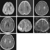
Patient 3 had steroid-induced IDDM and presented during induction. Initial strokelike event was associated with aphasia and right hemiparesis and hemisensory loss, which resolved. Second event occurred 8 weeks later and was associated with aphasia, left hemiparesis, and focal seizure, all of which resolved. A and B, DW image (A) and ADC map (B) at initial presentation show large areas of restricted diffusion in the centrum semiovale (arrows). Bilateral diffusion abnormalities were associated with unilateral hemimotor-sensory deficit. C and D, Axial FLAIR images obtained 8 weeks later shows extensive presumed demyelination in deep white matter. Note absence of abnormal signal intensity in the right parietal region. E and F, Axial DW image (E) and ADC map (F) show restricted diffusion in the right parietal cortex and subcortical white matter. Diffusion abnormalities were correlated with left hemiparesis and left focal seizure. Hyperintensity in the left anterior centrum semiovale (arrow in E) is due to T2 shine-through; ADC map shows a corresponding area of hyperintensity (arrow in F). G, Axial FLAIR image obatined 28 months later when the patient was asymptomatic shows more-extensive white matter abnormalities. Note absence of gliosis in the right parietal region.
F<sc>ig</sc> 4.
Fig 4.
Patient 4 presented with nonfluent aphasia and alternating fluctuating hemiparesis and left facial droop. A and B, Axial DW images show asymmetric regions of restricted diffusion in the anterior centrum semiovale that do not explain the motor neurologic deficits. C, Follow-up MR image obtained 39 months later shows minimal scattered demyelination. The patient was neurologically intact.
F<sc>ig</sc> 5.
Fig 5.
Patient 5 presented with nonfluent aphasia, mild right facial weakness, right hemiparesis, and left-arm sensory deficit. A, DW image shows a small area of restricted diffusion with the left precentral region (arrow) that does not fully explain the neurologic deficits. B, Axial FLAIR image obtained 13 months later shows no signal-intensity abnormality in the left precentral region. C, Image shows small areas of presumed demyelination in the centrum semiovale. The patient was neurologically intact.

Similar articles

Cited by

References

    1. Winick NJ, Bowman WP, Kamen BA, et al. Unexpected acute neurologic toxicity in the treatment of children with acute lymphoblastic leukemia. J Natl Cancer Inst 1992;84:252–256 - PubMed
    1. Mahoney DH, Shuster JJ, Nitschke R, et al. Acute neurotoxicity in children with B-precursor acute lymphoid leukemia: an association between intermediate–dose intravenous methotrexate and intrathecal triple therapy: a Pediatric Oncology Group study. J Clin Oncol 1998;16:1712–1722 - PubMed
    1. Winick N, Shuster JJ, Bowman WP, et al. Intensive oral methotrexate protects against lymphoid marrow relapse in childhood B-precursor acute lymphoblastic leukemia. J Clin Oncol 1996;14:2803–2811 - PubMed
    1. Rubnitz JE, Relling MV, Harrison, et al. Transient leukoencephalopathy following high dose methotrexate treatment in childhood acute lymphocytic leukemia. Leukemia 1998;12:1176–1181 - PubMed
    1. Rubinstein LJ, Herman MM, Long TF, Wilbur JR. Disseminated necrotizing leukoencephalopathy: a complication of treated central nervous system leukemia and lymphoma. Cancer 1975;35:291–305 - PubMed

MeSH terms

Substances

LinkOut - more resources