

Evaluation of cortical atrophy between progressive supranuclear palsy and corticobasal degeneration by hemispheric surface display of MR images
- PMID: 15569735
- PMCID: PMC8148711
Evaluation of cortical atrophy between progressive supranuclear palsy and corticobasal degeneration by hemispheric surface display of MR images
Abstract
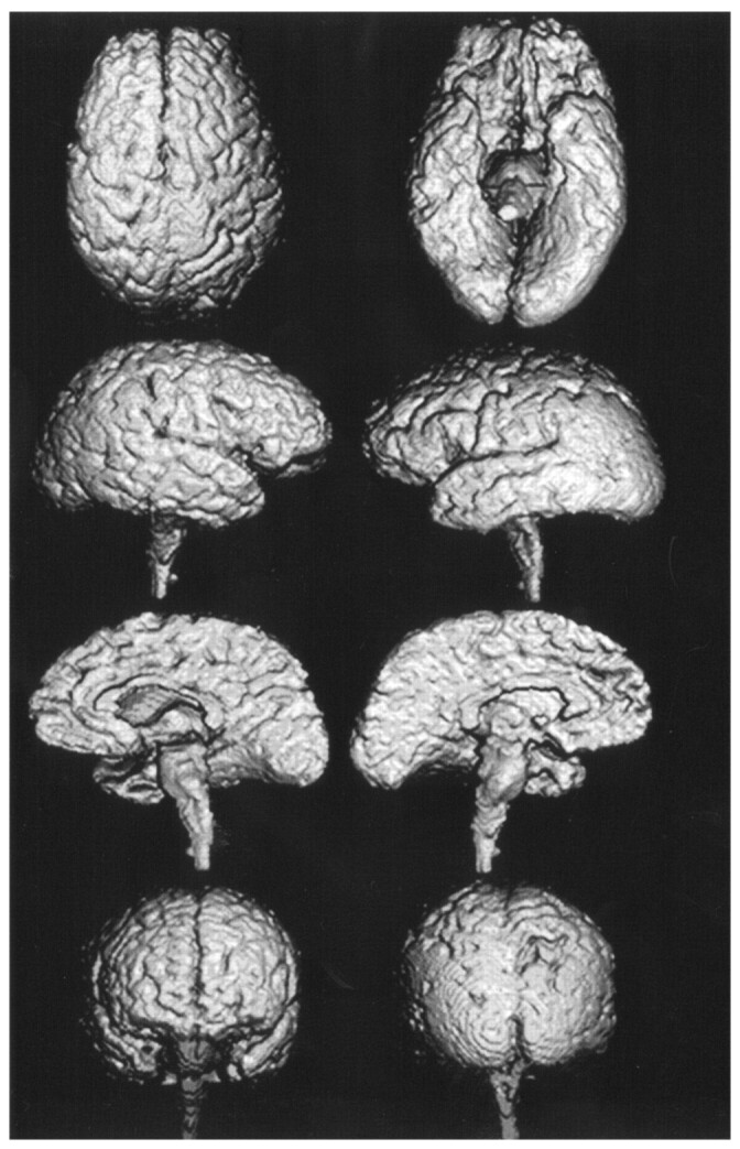
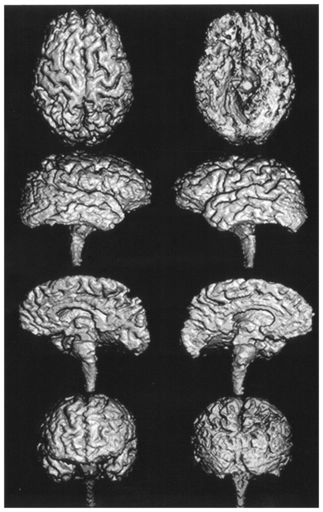
Background and purpose: Three-dimensional imaging and hemispheric volumetry are useful for the assessment of degenerative cortical atrophy. Our purpose was to determine the features of cortical atrophy in progressive supranuclear palsy (PSP) and corticobasal degeneration (CBD) by means of a hemispheric surface display generated with MR images.
Methods: The extent of cortical atrophy was evaluated with automated MR hemispheric surface display and volumetry in 19 patients with PSP and 19 with CBD.
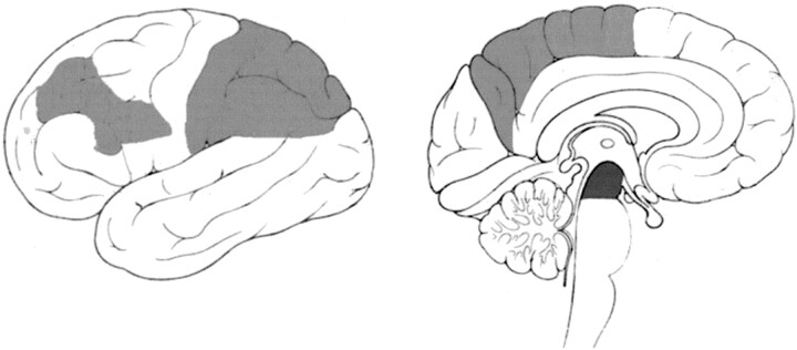
Results: Most cortical regions were less atrophic in PSP than in CBD. The parietal lobe, paracentral regions, anterior middle frontal lobe, and posterior inferior frontal lobe were significantly more atrophic in CBD than in PSP, whereas the brainstem was significantly more atrophic in PSP. The mean hemisphere-to-intracranial volume ratio was significantly greater in patients with PSP (74.5%) than in those with CBD (71.4%), whereas the mean brainstem-to-intracranial volume ratio was significantly smaller in PSP (1.4%) than in CBD (1.6%). Asymmetry of hemispheric volume was significantly larger in the CBD group than in the PSP group.
Conclusion: Hemispheric surface display and volumetry are generally helpful and especially useful for the differentiation of PSP and CBD.
Figures
References
-
- Dickson DW. Neuropathologic differentiation of progressive supranuclear palsy and corticobasal degeneration. J Neurol 1999;246:II6–II15 - PubMed
-
- Di Maria E, Tabaton M, Vigo T, et al. Corticobasal degeneration shares a common genetic background with progressive supranuclear palsy. Ann Neurol 2000;47:374–377 - PubMed
-
- Yagishita A, Oda M. Progressive supranuclear palsy: MRI and pathological findings [Suppl]. Neuroradiology 1996;1:S60–S66 - PubMed
-
- Grisoli M, Fetoni V, Savoiardo M, Girotti F, Bruzzone MG. MRI in corticobasal degeneration. Eur J Neurol 1995;2:547–552 - PubMed
-
- Hauser RA, Murtaugh FR, Akhter K, Gold M, Olanow CW. Magnetic resonance imaging of corticobasal degeneration. J Neuroimaging 1996;6:222–226 - PubMed
MeSH terms
LinkOut - more resources
Full Text Sources
Medical
Miscellaneous