

fMRI biomarker of early neuronal dysfunction in presymptomatic Huntington's Disease
- PMID: 15569736
- PMCID: PMC8148746
fMRI biomarker of early neuronal dysfunction in presymptomatic Huntington's Disease
Abstract
Background and purpose: Functional MR imaging (fMRI) has been used to probe basal ganglia function in people with presymptomatic Huntington's disease (pre-HD). A previous fMRI study in healthy individuals demonstrated activation of the basal ganglia during a time-discrimination task. The current study was designed to examine the relative sensitivity of fMRI compared with that of behavioral testing and morphometric measurements in detecting early neurodegenerative changes related to Huntington's disease (HD).
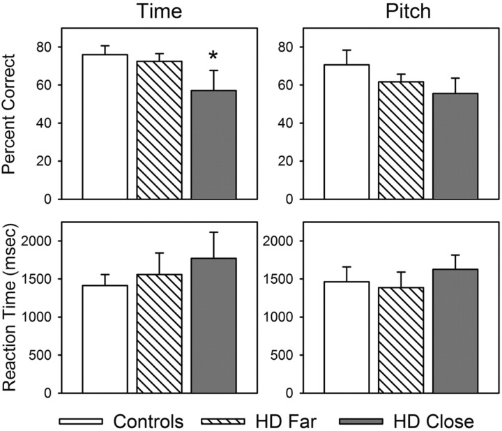
Methods: Pre-HD participants were assigned to two groups based on estimated years to diagnosis of manifest disease: close <12 years and far >or=12 years. Age at disease onset was estimated using a regression equation based on the number of trinucleotide CAG repeats. The time-discrimination task required participants to determine whether a specified interval was shorter or longer than a standard interval of 1200 milliseconds.
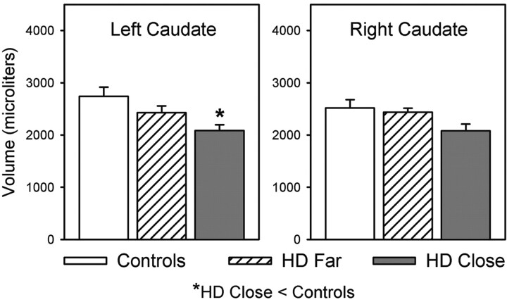
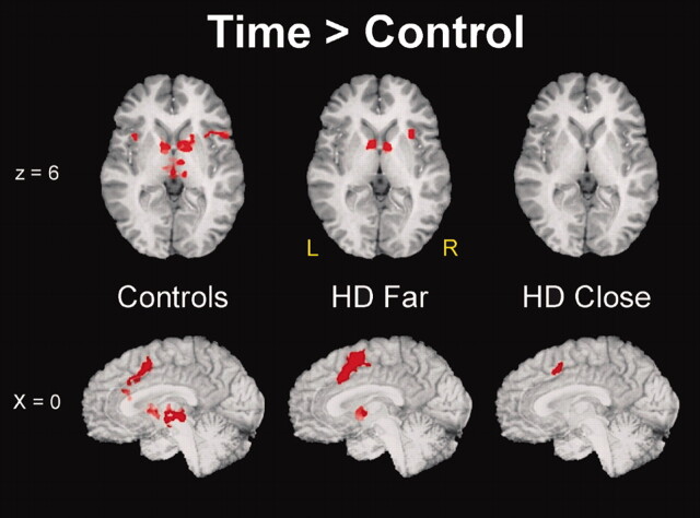
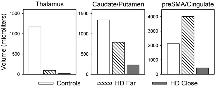
Results: Participants in the close group performed more poorly on the time-task discrimination than did control subjects; however, no differences were observed between far participants and control subjects. Similarly, close participants had reduced bilateral caudate volume relative to that of control subjects, whereas far participants did not. On functional imaging, close participants had significantly less activation in subcortical regions (caudate, thalamus) than control subjects; far participants had an intermediate degree of activation. In contrast, far participants had hyperactivation in medial hemispheric structures (anterior cingulate, pre-supplementary motor area) relative to close and control subjects.
Conclusion: Hyperactivation of medial prefrontal regions compensated for reduced subcortical participation during time discrimination in pre-HD. This pattern of brain activation may represent an early neurobiologic marker of neuronal dysfunction.
Figures
References
-
- The Huntington’s Disease Collaborative Research Group: a novel gene containing a trinucleotide repeat that is expanded and unstable on Huntington’s disease chromosomes. Cell 1993;72:971–983 - PubMed
-
- Vonsattel JP, Myers RH, Stevens TJ, Ferrante RJ, Bird ED, Richardson EP Jr. Neuropathological classification of Huntington’s disease. J Neuropathol Exp Neurol 1985;44:559–577 - PubMed
-
- Myers RH, Vonsattel JP, Stevens TJ, et al. Clinical and neuropathologic assessment of severity in Huntington’s disease. Neurology 1988;38:341–347 - PubMed
-
- Wagster MV, Hedreen JC, Peyser CE, Folstein SE, Ross CA. Selective loss of [3H]kainic acid and [3H]AMPA binding in layer VI of frontal cortex in Huntington’s disease. Exp Neurol 1994;127:70–75 - PubMed
-
- Sotrel A, Paskevich PA, Kiely DK, Bird ED, Williams RS, Myers RH. Morphometric analysis of the prefrontal cortex in Huntington’s disease. Neurology 1991;41:1117–1123 - PubMed
Publication types
MeSH terms
Grants and funding
LinkOut - more resources
Full Text Sources
Medical