Case Reports
. 2004 Nov-Dec;25(10):1722-9.

CNS MR and CT findings associated with a clinical presentation of herpetic acute retinal necrosis and herpetic retrobulbar optic neuritis: five HIV-infected and one non-infected patients

Affiliations
Case Reports

CNS MR and CT findings associated with a clinical presentation of herpetic acute retinal necrosis and herpetic retrobulbar optic neuritis: five HIV-infected and one non-infected patients

Robert J Bert et al. AJNR Am J Neuroradiol. 2004 Nov-Dec.

Abstract

Introduction: This report demonstrates the spectrum of central nervous system (CNS) abnormalities observed on MR imaging and CT studies in 6 patients with clinical or pathologic diagnoses of acute retinal necrosis (ARN) and retrobulbar optic neuritis (RBON-H) resulting from Herpes Zoster Virus and Cytomegalovirus. We discuss the etiologic and pathophysiologic implications regarding these findings.

Methods: Standard MR imaging sequences of the whole brain and selected high-resolution images of the orbits and globes, from 6 patients, were reviewed by three neuroradiologists for consensus interpretation of the findings. Special sequences augmenting disease were obtained in individual cases. Axial CT images were obtained from two patients using 5mm sequential slices.

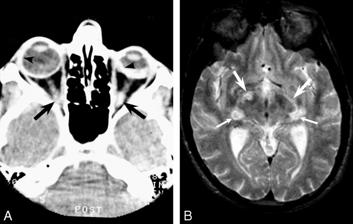
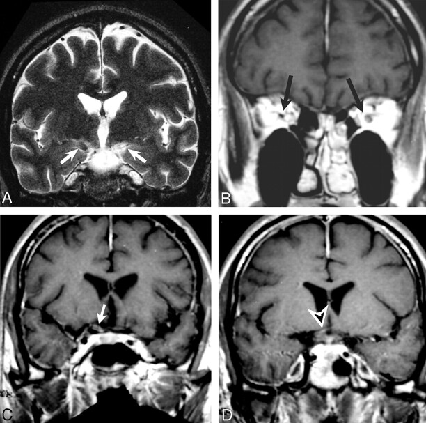
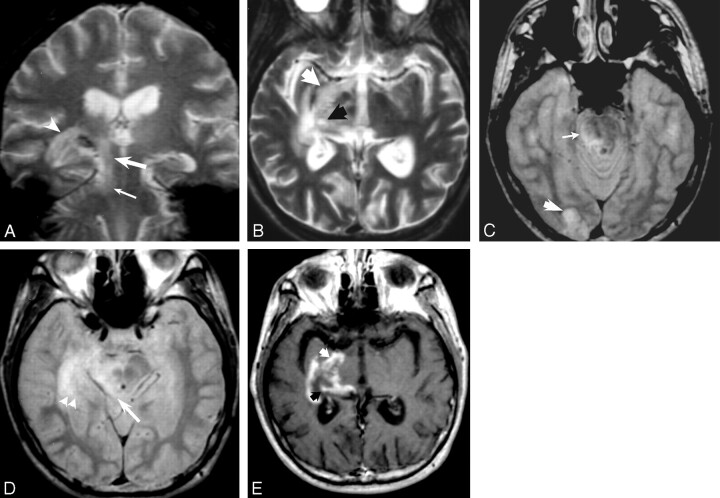
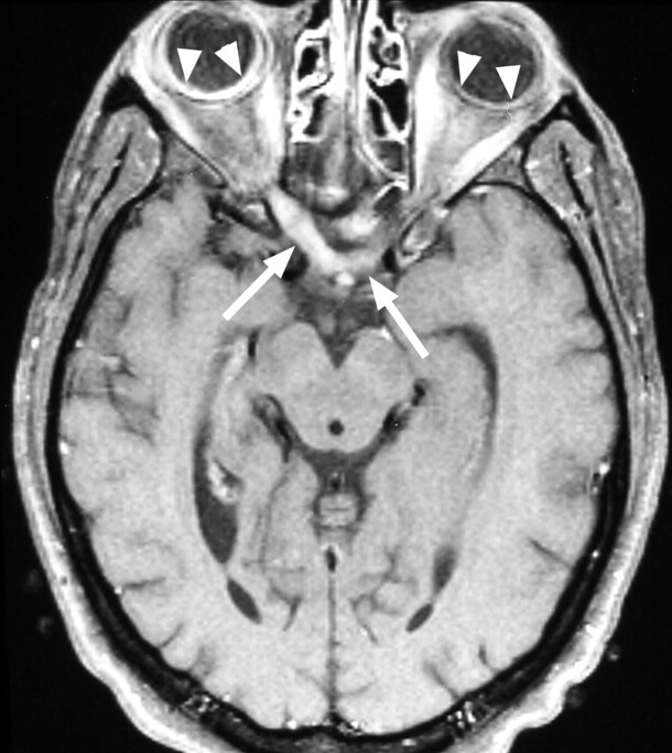
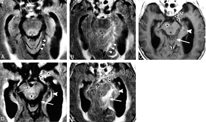
Results: MR imaging findings showed both T2 signal brightening and contrast enhancement in one or both optic nerves, optic tracts and lateral geniculate bodies, as well as the postsynaptic optic radiations and optic cortex. Similar findings were observed in the superior colliculus, lateral midbrain and cerebellum, with multiple potential etiologic possibilities regarding pathways of dissemination. Low T2* signal (indicating magnetic field susceptibility effects) and CT hyperdensity, consistent with prior hemorrhage, were also observed in the optic tracts, optic radiations and lateral geniculate bodies. Post-contrast enhancement was observed in the meninges and Meckle's cave in one HIV negative patient.

Conclusion: These cases demonstrate CNS imaging findings associated with RBON that are temporally-related to ARN. They support the hypothesis that RBON can either precede or follow ARN and implicate transneuronal, transsynaptic and/or transcerebrospinal fluid viral spread by the herpetic family.


Figures

F<sc>ig</sc> 1.
Fig 1.
Images from 1988 obtained in a patient with AIDS with CMV-induced ARN preceding retrobulbar optic neuritis. A, CT scan shows a shrunken left globe with bilateral avidly enhancing sclera (arrowheads) and enhancement of both optic nerves (arrows). B, T2-weighted image shows increased signal intensity in the optic tracts (top arrows) and geniculate bodies (bottom arrows).
F<sc>ig</sc> 2.
Fig 2.
MR images obtained in patient 2. A, T2-weighted coronal image shows both increased (left) and decreased (right) signal in the optic tracks (arrows). B, Contrast-enhanced T1-weighted image shows enhancement of the left optic nerve and low signal intensity in the right optic nerve (arrows). C and D, Contrast-enhanced T1-weighted images show enhancement of the right optic tract (arrow in C) and chiasm (arrowhead in D).
F<sc>ig</sc> 3.
Fig 3.
Images obtained in patient 3 with HZV-induced progressive outer retinal necrosis preceding retrobulbar optic neuritis, CNS extension, and eventual demise. A, Coronal T2-weighted gradient-echo image suggests hemorrhage in the right lateral geniculate body, with increased signal intensity involving the optic radiation through the temporal lobe (arrowhead) and extending caudad along the lateral midbrain (large arrow) to the cerebellum (small arrow). B, Axial T2-weighted turbo spin-echo image shows increased signal intensity along the right optic tract (white arrow) and lateral geniculate body (black arrow). C and D, Proton density–weighted images demonstrate abnormally high signal intensity along the right hemipons (thin arrow in C), right midbrain, and superior colliculus (arrow in D), occipital (visual) cortex (thick arrow in C), and optic radiation in the temporal lobe (arrowheads in D). E, T1-weighted contrast-enhanced image shows enhancement in similar regions: optic tract (white arrow) and lateral geniculate body (black arrow). Postmortem immunofluorescence stains (not shown) demonstrated HZV in these areas.
F<sc>ig</sc> 4.
Fig 4.
Patient 4 with HZV-induced progressive outer retinal necrosis and retrobulbar optic neuritis after herpes ophthalmicus. T1-weighted contrast-enhanced image shows enhancement of the optic nerves (right distal, left proximal) and optic chiasm (arrows). Compare the thickened, avidly enhancing right chorioretina with the normally enhancing left retina (arrowheads).
F<sc>ig</sc> 5.
Fig 5.
Axial images obtained in elderly HIV-negative woman with HZV ophthalmicus preceding retrobulbar optic neuritis and mental status changes. A and B, Nonenhanced and enhanced T2-weighted FLAIR images at the level of the pons show minimal abnormal high signal intensity in A and avid enhancement in B in the left ambient cistern, supracerebellar cistern, cerebellar vermis and adjacent subarachnoid space along the tentorium (arrowheads). CE, Axial images at the level of the midbrain. Contrast-enhanced conventional T1-weighted image in C shows minimal enhancement. Nonenhanced (D) and enhanced (E) T2-weighted FLAIR images show avid enhancement of the optic nerves, chiasm, tracts, and pericavernous region (black arrowheads), right lateral geniculate body (white arrowhead), interpeduncular cistern (black arrow), and supracerebellar cistern (white arrow).

Similar articles

Cited by

References

    1. Litoff D, Catalano RA. Herpes zoster optic neuritis in human immunodeficiency virus infection. Arch Ophthalmol 1990;108:782–783 - PubMed
    1. Selbst RG, Selhorst JB., Harbison JW, Myer EC. Parainfectious optic neuritis: report and review following varicella. Arch Neurol 1983;40:347–350 - PubMed
    1. Kuppermann BD, Quiceno JI, Wiley C, et al. Clinical and histopathologic study of varicella zoster virus retinitis in patients with the acquired immunodeficiency syndrome. Am J Ophthalmol 1994;188:589–600 - PubMed
    1. Mansour AM. Neuro-ophthalmic findings in acquired immunodeficiency syndrome. J Clin Neuro-Ophthalmol 1990;10:167–174 - PubMed
    1. Garweg J, Bohnke M. Varicella-zoster virus is strongly associated with atypical necrotizing herpetic retinopathies. Clin Infect Dis 1997;24:603–608 - PubMed

Publication types

MeSH terms