

Blood brain-barrier disruption of nonionic iodinated contrast medium following coil embolization of a ruptured intracerebral aneurysm
- PMID: 15569746
- PMCID: PMC8148708
Blood brain-barrier disruption of nonionic iodinated contrast medium following coil embolization of a ruptured intracerebral aneurysm
Erratum in
- AJNR Am J Neuroradiol. 2005 Jan;26(1):203
Abstract
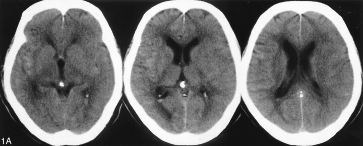
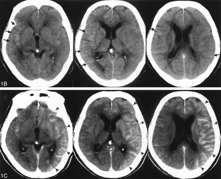
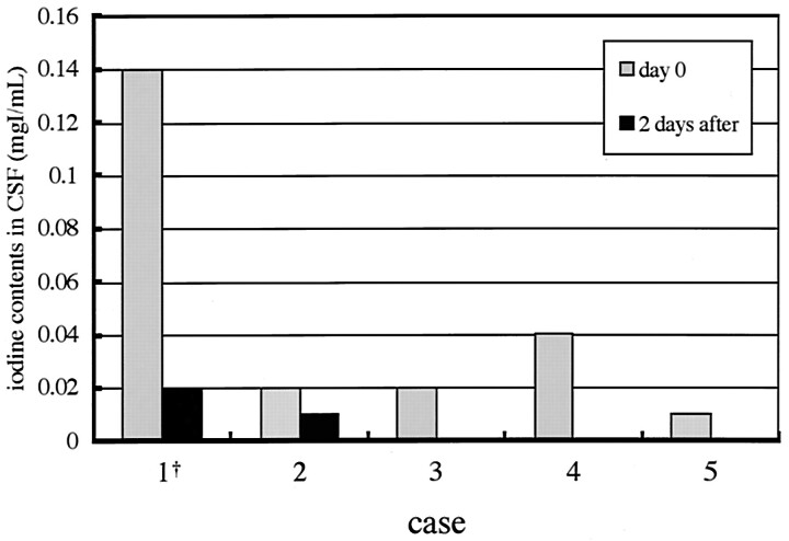
Few reports of temporary disruption of the blood-brain barrier (BBB) following neurointerventional procedures, presumably caused by nonionic radiographic contrast medium (CM), exist in the literature. We described such a case in a 72-year-old man presenting with acute subarachnoid hemorrhage, who underwent coil embolization of a ruptured anterior communicating artery complex aneurysm. At the time of his follow-up CT examination, a large amount of iodine was found in the cerebrospinal fluid (CSF). Because of this experience, the iodine concentration in the CSF of five other patients who also underwent an intracranial endovascular procedure was measured. It was concluded that this increased iodine might have been caused by temporary leakage or breakdown of the BBB. Even if the total amount of CM may not be excessive, the disproportionately high concentration injected into a single vascular territory may pose a unique set of variables increasing the risk of BBB disruption.
Figures
References
-
- Velden J, Milz P, Winkler F, et al. Nonionic contrast neurotoxicity after coronary angiography mimicking subarachnoid hemorrhage. Eur Neurol 2003;49:249–251 - PubMed
-
- Sticherling C, Berkfield J, Auch-Schwelk W, Lanfermann H. Transient bilateral cortical blindness after coronary angiography. Lancet 1998;351:570. - PubMed
-
- Earnest FIV, Forbes G, Sandok BA, et al. Complications of cerebral angiography: prospective assessment of risk. AJNR Am J Neuroradiol 1983;4:1191–1197 - PubMed
-
- Numaguchi Y, Fleming MS, Hasuo K, et al. Blood-brain barrier disruption due to cerebral arteriography: CT findings. J Comput Assist Tomogr 1984;8:936–939 - PubMed
Publication types
MeSH terms
Substances
LinkOut - more resources
Full Text Sources
Medical