

Retroclival ecchordosis physaliphora: MR imaging and review of the literature
- PMID: 15569763
- PMCID: PMC8148737
Retroclival ecchordosis physaliphora: MR imaging and review of the literature
Abstract
Background and purpose: Ecchordosis physaliphora (EP), found in about 2% of autopsies, is a clinically inconspicuous notochordal remnant appearing at the dorsal wall of the clivus. To our knowledge, a systematic review of its MR features does not exist. The aim of this study was to describe the MR imaging findings of incidentally found retroclival EP with special respect to its differentiation from intradural chordomas.
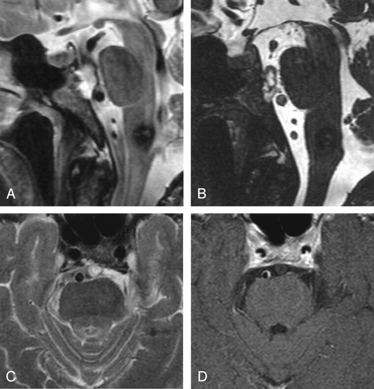
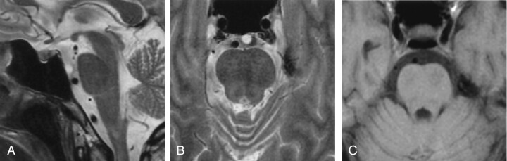
Methods: We reviewed 300 consecutive 1.5-T MR imaging studies that included thin-section transverse T2-weighted images of the skull base for the presence of a retroclival EP. In cases in which an EP was identified, two neuroradiologists observed MR signal intensity characteristics, contrast enhancement, size, form, stalk of EP, and signal intensity changes of the adjacent clivus.
Results: Five cases with retroclival EP were found (incidence, 1.7%). In all cases, the ecchordoses was hyperintense on T2-weighted images and hypointense on T1-weighted images. Contrary to the reported findings in chordomas, none of the lesions showed contrast enhancement. In four cases, there were signal intensity changes in the adjacent clivus. A stalklike connection between clivus and EP was seen in three patients.
Conclusion: Because of the benign character of EP and the difficulties in its histopathologic differentiation from chordomas, precise knowledge of the radiologic characteristics of EP is important. On the basis of these five cases and a review of literature, contrast enhancement and the presence of clinical symptoms seem to be highly reliable parameters in the differential diagnosis of intradural chordoma and EP.
Figures
References
-
- Wolfe JT III, Scheithauer BW. “Intradural chordoma” or “giant ecchordosis physaliphora”? Report of two cases. Clin Neuropath 1987;6:98–103 - PubMed
-
- Lantos PL, Louis DN, Rosenblum MK, Kleihues P. Tumours of the nervous system. In: Graham DI, Lantos PL, eds. Greenfield’s Neuropathology. 7th ed. Vol. 2. London: Arnold;2002;767–1052
-
- Rodriguez L, Colina J, Lopez J, Molina O, Cardozo J. Intradural prepontine growth: giant ecchordosis physaliphora or extraosseous chordoma? Neuropathology 1999;19:336–340
-
- Toda H, Kondo A, Iwaski K. Neuroradiological characteristics of ecchordosis physaliphora: case report and review of the literature. J Neurosurg 1998;89:830–834 - PubMed
Publication types
MeSH terms
LinkOut - more resources
Full Text Sources
Medical