

Costimulatory B7-H1 in renal cell carcinoma patients: Indicator of tumor aggressiveness and potential therapeutic target
- PMID: 15569934
- PMCID: PMC534606
- DOI: 10.1073/pnas.0406351101
Costimulatory B7-H1 in renal cell carcinoma patients: Indicator of tumor aggressiveness and potential therapeutic target
Abstract
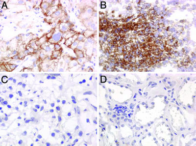
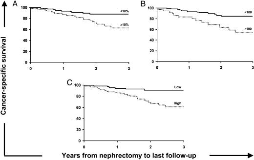
Expression of B7-H1, a costimulating glycoprotein in the B7 family, is normally restricted to macrophage-lineage cells, providing a potential costimulatory signal source for regulation of T cell activation. In contrast, aberrant expression of B7-H1 by tumor cells has been implicated in impairment of T cell function and survival, resulting in defective host antitumoral immunity. The relationship between tumor-associated B7-H1 and clinical cancer progression is unknown. Herein, we report B7-H1 expression by both renal cell carcinoma (RCC) tumors of the kidney and RCC tumor-infiltrating lymphocytes. In addition, our analysis of 196 clinical specimens reveals that patients harboring high intratumoral expression levels of B7-H1, contributed by tumor cells alone, lymphocytes alone, or tumor and/or lymphocytes combined, exhibit aggressive tumors and are at markedly increased risk of death from RCC. In fact, patients with high tumor and/or lymphocyte B7-H1 levels are 4.5 times more likely to die from their cancer than patients exhibiting low levels of B7-H1 expression (risk ratio 4.53; 95% confidence interval 1.94-10.56; P < 0.001.) Thus, our study suggests a previously undescribed mechanism whereby RCC may impair host immunity to foster tumor progression. B7-H1 may prove useful as a prognostic variable for RCC patients both pre- and posttreatment. In addition, B7-H1 may represent a promising target to facilitate more favorable responses in patients who require immunotherapy for treatment of advanced RCC.
Figures
References
-
- Dong, H., Zhu, G., Tamada, K. & Chen L. (1999) Nat. Med. 5, 1365–1369. - PubMed
-
- Dong, H., Strome, S.-E., Salomao, D.-R., Tamura, H., Hirano, F., Flies, D.-B., Roche, P.-C., Lu, J., Zhu, G., Tamada, K., et al. (2002) Nat. Med. 8, 793–800. - PubMed
-
- Strome, S.-E. & Chen, L. (2004) Curr. Treat. Options Oncol. 5, 27–33. - PubMed
-
- Dong, H. & Chen, L. (2003) J. Mol. Med. 81, 281–287. - PubMed
-
- Strome, S.-E., Dong, H., Tamura, H., Voss, S.-G., Flies, D.-B., Tamada, K., Salomao, D., Cheville, J.-C, Hirano, F., Lin, W., et al. (2003) Cancer Res. 63, 6501–6505. - PubMed
MeSH terms
Substances
LinkOut - more resources
Full Text Sources
Other Literature Sources
Medical
Research Materials
