. 2004 Dec 7;101(49):17294-9.
doi: 10.1073/pnas.0404743101. Epub 2004 Nov 29.

Targeted disruption of luteinizing hormone beta-subunit leads to hypogonadism, defects in gonadal steroidogenesis, and infertility

Affiliations

Targeted disruption of luteinizing hormone beta-subunit leads to hypogonadism, defects in gonadal steroidogenesis, and infertility

Xiaoping Ma et al. Proc Natl Acad Sci U S A. .

Abstract

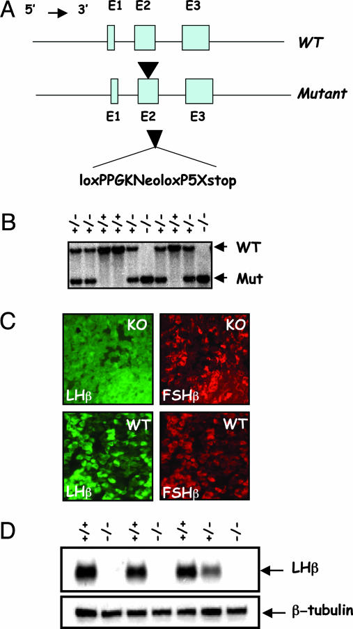
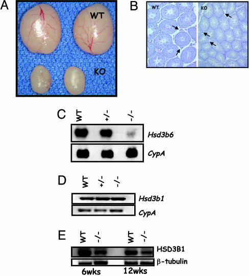
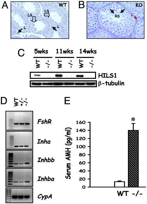
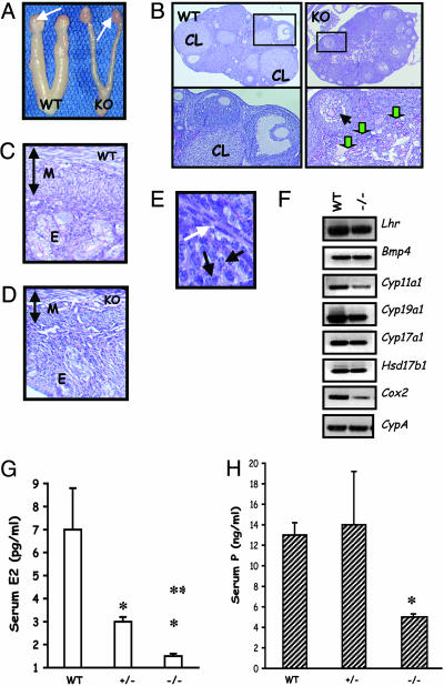
Luteinizing hormone (LH) and follicle-stimulating hormone (FSH) act on gonadal cells to promote steroidogenesis and gametogenesis. Clarifying the in vivo roles of LH and FSH permits a feasible approach to contraception involving selective blockade of gonadotropin action. One way to address these physiologically important problems is to generate mice with an isolated LH deficiency and compare them with existing FSH loss-of-function mice. To model human reproductive disorders involving loss of LH function and to define LH-responsive genes, we produced knockout mice lacking the hormone-specific LHbeta-subunit. LHbeta-null mice are viable but demonstrate postnatal defects in gonadal growth and function resulting in infertility. Mutant males have decreased testes size, prominent Leydig cell hypoplasia, defects in expression of genes encoding steroid biosynthesis pathway enzymes, and reduced testosterone levels. Furthermore, spermatogenesis is blocked at the round spermatid stage, causing a total absence of the elongated spermatids. Mutant female mice are hypogonadal and demonstrate decreased levels of serum estradiol and progesterone. Ovarian histology demonstrates normal thecal layer, defects in folliculogenesis including many degenerating antral follicles, and absence of corpora lutea. The defects in both sexes are not secondary to aberrant FSH regulation, because FSH levels were unaffected in null mice. Finally, both male and female null mice can be pharmacologically rescued by exogenous human chorionic gonadotropin, indicating that LH-responsiveness of the target cells is not irreversibly lost. Thus, LHbeta null mice represent a model to study the consequences of an isolated deficiency of LH ligand in reproduction, while retaining normal LH-responsiveness in target cells.


Figures

Fig. 1.
Fig. 1.
Gene targeting at the LHβ locus. (A) A phosphoglycerate kinase-neomycin expression cassette was engineered to contain a 5′ loxP sequence and on the 3′ side another loxP sequence followed by five stop codons. This cassette was inserted into exon 2 of the mouse LHβ gene in ES cells by homologous recombination (arrowhead). The floxed phosphoglycerate kinase-neomycin cassette was flanked by 6.3 kb of 5′ and 6 kb of 3′ LHβ gene sequences originally cloned from a 129S6/SvEv mouse genomic library. (B) Southern blot shows WT and mutant alleles identified by their size (7.3 and. 3.4 kb, respectively) to distinguish the genotypes of mice. (C) Dual immunofluorescence of pituitary sections shows that LHβ-specific staining (labeled green) was absent in a pituitary section from the null mouse (KO) compared with that in control (WT). FSHβ-specific staining (in red) is seen in both control and null mice. (D) Immunoblot of pituitary proteins shows the presence of LHβ-reactive band only in control pituitary extracts (+/+ and +/–) but not in that of the null (–/–) mice, thus confirming that we engineered a null mutation at the LHβ locus. Because heterodimer assembly confers biological activity to glycoprotein hormones, absence of LHβ-subunit leads to LH deficiency in these null mice.
Fig. 2.
Fig. 2.
Male reproductive phenotypes of LHβ knockout mice. (A) Morphology of testes from adult (6 weeks) control (WT) and null (KO) male mice. Note the reduction in size due to the absence of LHβ.(B Left) Low-power histology shows many normal Leydig cells in the WT testis (arrows), whereas the mutant testis (Right) shows sparse interstitium and small Leydig cells (arrows). Note the reduction in tubule size in KO testis section. (Photographed at ×5 magnification.) (C Upper) Northern blot analysis indicates that expression of adult Leydig cell specific 3β-hydroxysteroid dehydrogenase type VI (Hsd3b6) is suppressed in the null (–/–) testes. (Lower) Hybridization with a cyclophilin (CypA) probe confirms that an equal amount of RNA was loaded in each lane. (D and E) Expression of 3β-hydroxysteroid dehydrogenase type I in the mutant testes assessed by RT-PCR assay (D) and Western blot analysis (E) shows that although the steady-state levels of RNA were not changed, the protein is reduced in the null (–/–) testes. CypA amplification was used as a control for RT-PCR assay, and β-tubulin expression was used as an internal control for Western blot analysis.
Fig. 3.
Fig. 3.
Spermatogenesis arrest and Sertoli cell marker expression. (A and B) High-power histology of WT (A) and KO (B) testis sections shows Leydig cells (arrows, L). In contrast to many late-stage spermatids (open arrows, LS) and abundant sperm in the lumen of WT testis (A), spermatogenesis proceeds up to the round-spermatid stage (arrows, RS) in the KO testis (B). Note that other stages of spermatogenesis grossly appear normal in the KO testis section (B). (Magnification: A, ×10; B, ×20.) (C) Western blot analysis shows that HILS1, a late spermatid stage-specific marker, is not expressed in the mutant testis and confirms the block in spermatogenesis at the transition between round to elongated spermatid step. (D) RT-PCR analysis of testis cDNAs identifies that expression of Sertoli cell markers, including activin subunits, Inhba and Inhbb was increased, whereas that of FSH receptor and inhibin-α in the mutant testes remained unchanged. (E) Assay of serum levels of AMH indicates that this Sertoli cell marker was elevated in the mutant (–/–). *, P < 0.05 (n = 4).
Fig. 4.
Fig. 4.
Female reproductive phenotypes. (A) Morphological analysis of the female reproductive tract indicates hypoplastic uteri and ovaries in the mutant (KO, Right) compared with those in a WT control (Left) at 9 weeks. White arrows denote ovaries. (B) Low-power histology of the ovary shows many corpora lutea (CL) in the WT (Left) but not in the mutant (KO, Right). Multiple follicles at different stages are present in the mutant section, but preovualtory follicles are not present (Right Upper). (Left Lower) A magnified region (rectangle) of the WT section that contains CL, an antral follicle with a healthy oocyte. (Right Lower) A magnified region (rectangle) of the KO ovary section with a follicle containing a collapsed oocyte (black arrow) and many degenerating follicles with remnants of zona pellucida (green arrows). (Magnification: ×5.) (C and D) Histology of a 9-week-old null (KO, D) mouse uterus shows a thin myometrium (M) and hypoplastic endometrium (E), compared with that from a control (WT) mouse (C). The vertical doublehead arrows indicate the relative thickness of the myometrium in both cases. (E) High-power image of a follicle from KO ovary shows theca (white arrow) is formed in the absence of LH. Black arrows indicate granulosa cells for reference to the interior of the follicle. (Magnification: ×20.) (F) RT-PCR assay indicates that thecal cell markers LH receptor and BMP4 are expressed in the null (–/–) ovary. Many steroidogenic pathway enzymes and cycloxygenase 2 are suppressed in the null ovary in the absence of LH. Lhr, LH receptor; Bmp4, bone morphogenetic protein 4; Cyp11a1, P450 side chain cleavage enzyme11a1; Cyp19a1, cytochrome P450 aromatase; Cyp17a1, cytochrome P450 17-α hydroxylase; Hsd17b1, 17-β dehydrogenase type I; Cox2, cycloxygenase 2. (G and H) Consistent with the ovarian and uterine phenotype, serum estradiol (E2; G) and progesterone (P; H) are suppressed in the null mutants (–/–), but only E2 levels are suppressed in the heterozygous mutants (+/–). *, P < 0.05 vs. WT; **, P < 0.05 vs. ±.

References

    1. Pierce, J. G. & Parsons, T. F. (1981) Annu. Rev. Biochem. 50, 465–495. - PubMed
    1. Bousfield, G. R., Perry, W. M. & Ward, D. N. (1994) in The Physiology of Reproduction, eds. Knobil, E. & Neill, J. D. (Raven, New York), pp. 1749–1792.
    1. O'Shaughnessy, P. J., Baker, P., Sohnius, U., Haavisto, A. M., Charlton, H. M. & Huhtaniemi, I. (1998) Endocrinology 139, 1141–1146. - PubMed
    1. Pakarinen, P., Kimura, S., El-Gehani, F., Pelliniemi, L. J. & Huhtaniemi, I. (2002) Endocrinology 143, 4477–4482. - PubMed
    1. Saez, J. M., Perrard-Sapori, M. H., Chatelain, P. G., Tabone, E. & Rivarola, M. A. (1987) J. Steroid. Biochem. 27, 317–329. - PubMed

Publication types

Substances

LinkOut - more resources