

Does the novel PET/CT imaging modality impact on the treatment of patients with metastatic colorectal cancer of the liver?
- PMID: 15570208
- PMCID: PMC1356518
- DOI: 10.1097/01.sla.0000146145.69835.c5
Does the novel PET/CT imaging modality impact on the treatment of patients with metastatic colorectal cancer of the liver?
Abstract
Objective: To compare the diagnostic value of contrast-enhanced CT (ceCT) and 2-[18-F]-fluoro-2-deoxyglucose-PET/CT in patients with metastatic colorectal cancer to the liver.
Background: Despite preoperative evaluation with ceCT, the tumor load in patients with metastatic colorectal cancer to the liver is often underestimated. Positron emission tomography (PET) has been used in combination with the ceCT to improve identification of intra- and extrahepatic tumors in these patients. We compared ceCT and a novel fused PET/CT technique in patients evaluated for liver resection for metastatic colorectal cancer.
Methods: Patients evaluated for resection of liver metastases from colorectal cancer were entered into a prospective database. Each patient received a ceCT and a PET/CT, and both examinations were evaluated independently by a radiologist/nuclear medicine physician without the knowledge of the results of other diagnostic techniques. The sensitivity and the specificity of both tests regarding the detection of intrahepatic tumor load, extra/hepatic metastases, and local recurrence at the colorectal site were determined. The main end point of the study was to assess the impact of the PET/CT findings on the therapeutic strategy.
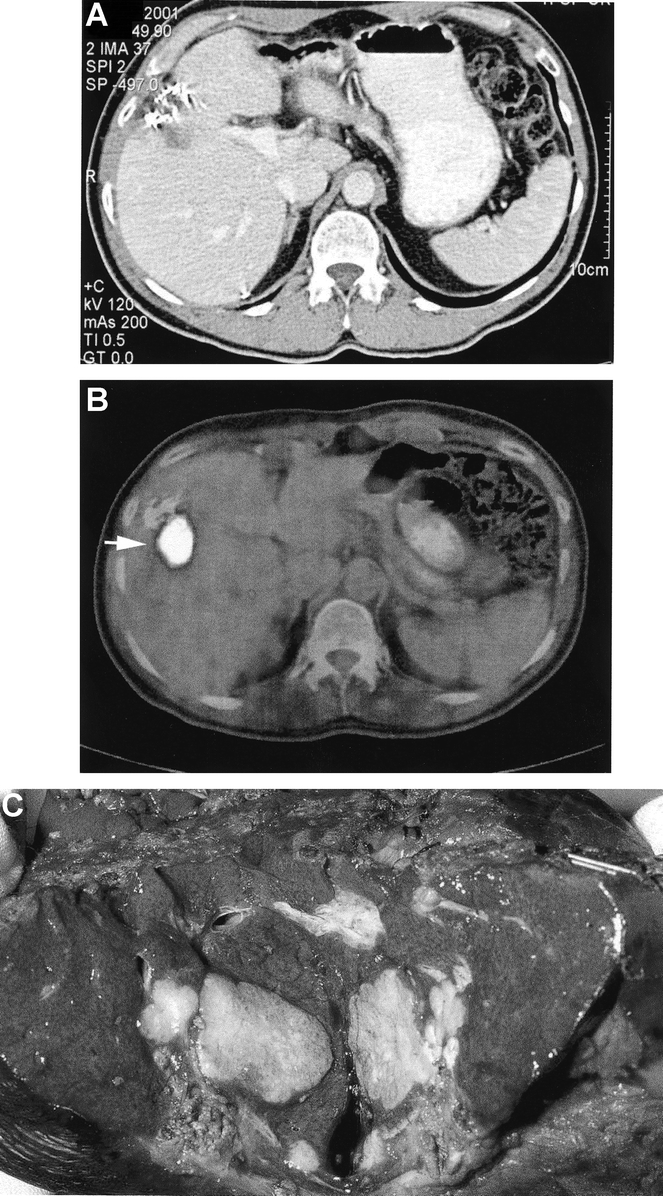
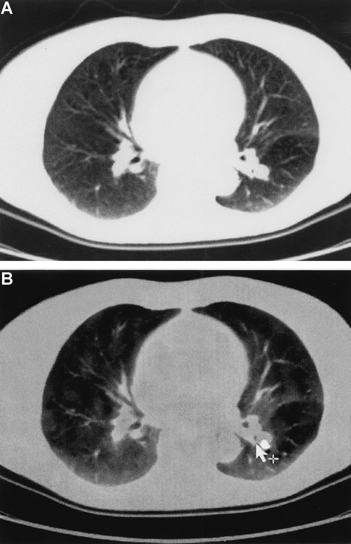
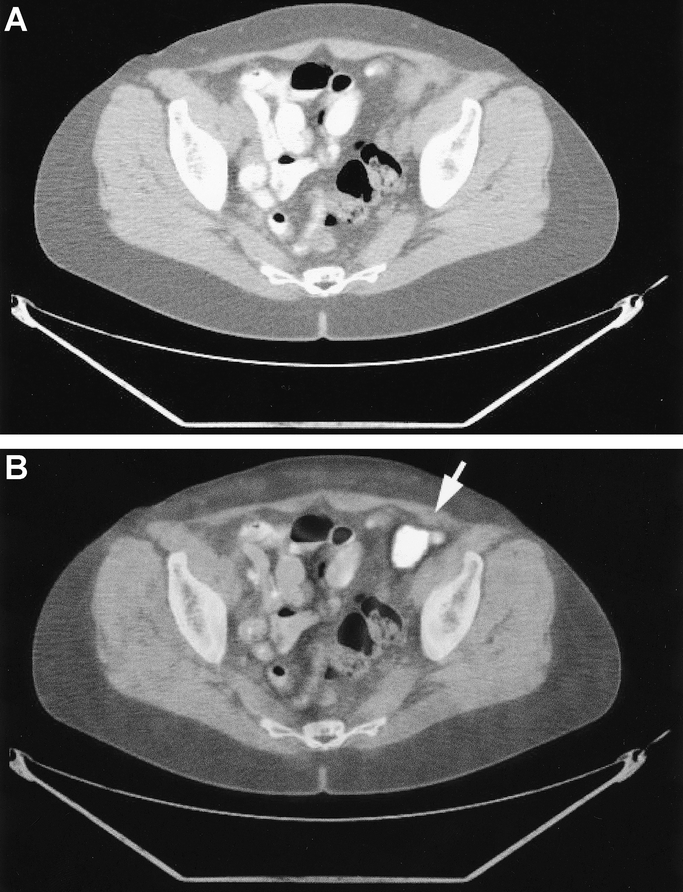
Results: Seventy-six patients with a median age of 63 years were included in the study. ceCT and PET/CT provided comparable findings for the detection of intrahepatic metastases with a sensitivity of 95% and 91%, respectively. However, PET/CT was superior in establishing the diagnosis of intrahepatic recurrences in patients with prior hepatectomy (specificity 50% vs. 100%, P = 0.04). Local recurrences at the primary colo-rectal resection site were detected by ceCT and PET/CT with a sensitivity of 53% and 93%, respectively (P = 0.03). Extrahepatic disease was missed in the ceCT in one third of the cases (sensitivity 64%), whereas PET/CT failed to detect extrahepatic lesions in only 11% of the cases (sensitivity 89%) (P = 0.02). New findings in the PET/CT resulted in a change in the therapeutic strategy in 21% of the patients.
Conclusion: PET/CT and ceCT provide similar information regarding hepatic metastases of colorectal cancer, whereas PET/CT is superior to ceCT for the detection of recurrent intrahepatic tumors after hepatectomy, extrahepatic metastases, and local recurrence at the site of the initial colorectal surgery. We now routinely perform PET/CT on all patients being evaluated for liver resection for metastatic colorectal cancer.
Figures
References
-
- Parker S, Tong T, Bolden S, et al. Cancer statistics. Cancer J Clin. 1997;47:5–27. - PubMed
-
- Selzner M, Clavien PA. Resection of liver tumors: Special emphasis on neoadjuvant and adjuvant therapy. In: Clavien PA, editor. Malignant Liver Tumors: Current and Emerging Therapies. Malden: Blackwell Science; 1999:137–149.
-
- Belghiti J, Hiramatsu K, Benoist S, et al. Seven hundred forty-seven hepatectomies in the 1990s: an update to evaluate the actual risk of liver resection. J Am Coll Surg. 2000;191:38–46. - PubMed
Publication types
MeSH terms
Substances
LinkOut - more resources
Full Text Sources
Medical
