Case Reports
. 2004 Nov 30;4(1):20.
doi: 10.1186/1471-2377-4-20.

Homozygosity for a missense mutation in the 67 kDa isoform of glutamate decarboxylase in a family with autosomal recessive spastic cerebral palsy: parallels with Stiff-Person Syndrome and other movement disorders

Affiliations
Case Reports

Homozygosity for a missense mutation in the 67 kDa isoform of glutamate decarboxylase in a family with autosomal recessive spastic cerebral palsy: parallels with Stiff-Person Syndrome and other movement disorders

Clare N Lynex et al. BMC Neurol. .

Abstract

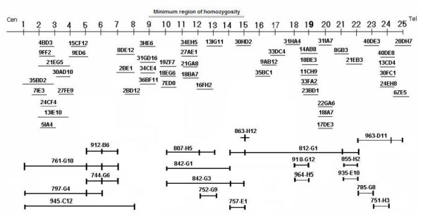
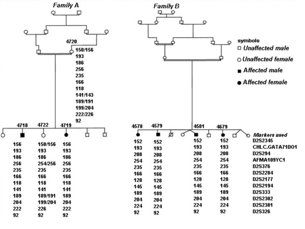
Background: Cerebral palsy (CP) is an heterogeneous group of neurological disorders of movement and/or posture, with an estimated incidence of 1 in 1000 live births. Non-progressive forms of symmetrical, spastic CP have been identified, which show a Mendelian autosomal recessive pattern of inheritance. We recently described the mapping of a recessive spastic CP locus to a 5 cM chromosomal region located at 2q24-31.1, in rare consanguineous families.

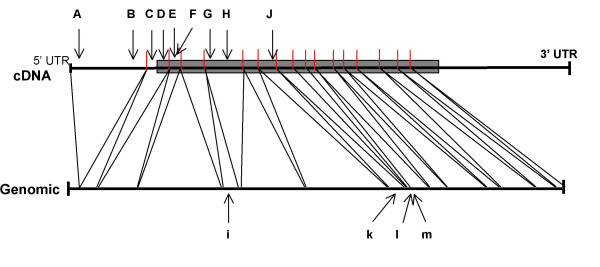
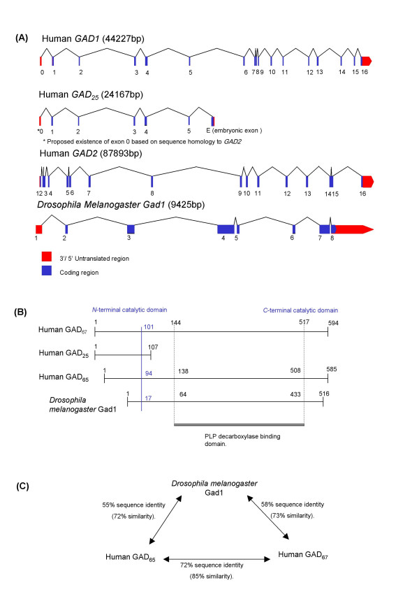
Methods: Here we present data that refine this locus to a 0.5 cM region, flanked by the microsatellite markers D2S2345 and D2S326. The minimal region contains the candidate gene GAD1, which encodes a glutamate decarboxylase isoform (GAD67), involved in conversion of the amino acid and excitatory neurotransmitter glutamate to the inhibitory neurotransmitter gamma-aminobutyric acid (GABA).

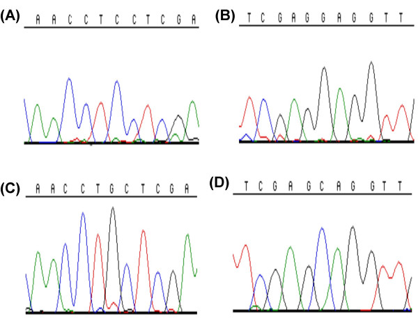
Results: A novel amino acid mis-sense mutation in GAD67 was detected, which segregated with CP in affected individuals.

Conclusions: This result is interesting because auto-antibodies to GAD67 and the more widely studied GAD65 homologue encoded by the GAD2 gene, are described in patients with Stiff-Person Syndrome (SPS), epilepsy, cerebellar ataxia and Batten disease. Further investigation seems merited of the possibility that variation in the GAD1 sequence, potentially affecting glutamate/GABA ratios, may underlie this form of spastic CP, given the presence of anti-GAD antibodies in SPS and the recognised excitotoxicity of glutamate in various contexts.


Figures

Figure 1
Figure 1
An integrated physical YAC contig spanning the human chromosome 2 spastic CP locus. This was constructed against a framework of microsatellite and STS markers, to incorporate the region of linkage identified by genotyping data. The positions of microsatellite and STS markers are represented numerically left to right from centromere to telomere. These loci 1–25 were mapped arbitrarily equi-distant onto the contig, in the following order: Cen-(1) D2S2157; (2) D2S382; (3) WI-18792; (4) D2S124; (5) D2S111; (6) D2S2384; (7) D2S2330; (8) D2S399; (9) D2S2345; (10) D2S294; (11) D2S2188; (12) D2S2284; (13) D2S2177; (14) D2S335; (15) D2S326; (16) D2S2381; (17) D2S2302; (18) D2S2307; (19) D2S2257; (20) D2S2314; (21) D2S138; (22) D2S148; (23) D2S2173; (24) D2S300; (25) D2S385-Tel.
Figure 2
Figure 2
Annotation of two pedigrees of spastic autosomal recessive CP families and corresponding linkage mapping data. The markers shown are those that demonstrate the minimal homozygous region between the affected individuals of both families.
Figure 3
Figure 3
Electropherograms of the sequence of the exon 1 SNP of GAD1 identified in the process of mutational analysis. (A) and (B) show the normal C variant in the forward and reverse directions, respectively. (C) and (D) show the alternative G variant in the forward and reverse directions, respectively. This variant was only found in affected individuals of family B. No heterozygous individuals were identified for this nucleotide variant.
Figure 4
Figure 4
An annotation of the distribution of single nucleotide substitutions identified in the open reading frame of GAD1. The approximate positions with respect to intron-exon of the open reading frame structure are illustrated. These were determined by sequencing of the probands in this study, from published data and from the NCBI collated database of SNPs. The letters refer to the SNPs listed in Table 4. Upper case letters refer to SNPs in the cDNA and lower case letters indicate SNPs in the genomic DNA. A: G(36)C, B: G(210)A, C: G(253)C, D: T(315)C, E: A(407)G, F: C(696)T, G: C(1506)T, H: C(1575)T, i: T(1625)G, J: C(1654)T, k: A(1659)G, l: G(1799)A, m: C(1899)A.
Figure 5
Figure 5
Three illustrations of the genomic, protein and comparative sequence homologies of the different species of GAD. (A) The genomic structures of GAD1/GAD25/GAD2 and Drosophila Gad1. (B) Comparative protein domain structures of GAD65/GAD25/GAD67 and Drosophila Gad1. (Numbers represent approximate amino acid residues). (C) Schematic illustrating the relative homology of the protein structures of GAD67/GAD65 and Drosophila Gad1.

References

    1. Eicher PS, Batshaw ML. Cerebral palsy. Pediatric Clinics of North America. 1993;40:537–551. - PubMed
    1. Gustavson KH, Hagberg B, Sanner G. Identical syndromes of cerebral palsy in the same family. Acta Paediatrica Scandinavica. 1969;58:330–340. - PubMed
    1. Kuban KCK, Leviton A. Cerebral palsy. New England Journal of Medicine. 1994;330:188–195. doi: 10.1056/NEJM199401203300308. - DOI - PubMed
    1. Volpe J. Neurology of the Newborn. 2. Philadelphia: WB Saunders; 1987.
    1. Gaffney G, Sellers S, Flavell V, Squier M, Johnson A. Case control study of intrapartum care, cerebral palsy and perinatal death. British Medical Journal. 1994;308:743–750. - PMC - PubMed

Publication types

Associated data