Review
. 2004 Dec;47(1-3):290-303.
doi: 10.1016/j.brainresrev.2004.08.002.

Role of connexin-based gap junction channels and hemichannels in ischemia-induced cell death in nervous tissue

Affiliations
Review

Role of connexin-based gap junction channels and hemichannels in ischemia-induced cell death in nervous tissue

Jorge E Contreras et al. Brain Res Brain Res Rev. 2004 Dec.

Abstract

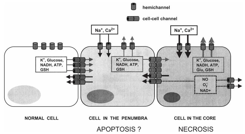
Gap junction channels and hemichannels formed of connexin subunits are found in most cell types in vertebrates. Gap junctions connect cells via channels not open to the extracellular space and permit the passage of ions and molecules of approximately 1 kDa. Single connexin hemichannels, which are connexin hexamers, are present in the surface membrane before docking with a hemichannel in an apposed membrane. Because of their high conductance and permeability in cell-cell channels, it had been thought that connexin hemichannels remained closed until docking to form a cell-cell channel. Now it is clear that at least some hemichannels can open to allow passage of molecules between the cytoplasm and extracellular space. Here we review evidence that gap junction channels may allow intercellular diffusion of necrotic or apoptotic signals, but may also allow diffusion of ions and substances from healthy to injured cells, thereby contributing to cell survival. Moreover, opening of gap junction hemichannels may exacerbate cell injury or mediate paracrine or autocrine signaling. In addition to the cell specific features of an ischemic insult, propagation of cell damage and death within affected tissues may be affected by expression and regulation of gap junction channels and hemichannels formed by connexins.


Figures

Fig. 1
Fig. 1
Scheme of the possible actions of connexin channels and hemichannels during and after ischemia. Hemichannels or gap junction channels with a dark center are open and are permeated by ions and small molecules. Those without a dark center are closed. Molecules required in normal metabolism (e.g., K+, glucose, NADH and reduced glutathione: GSH) can be released to the extracellular milieu through open hemichannels or can be transferred through gap junction channels from healthier cells (normal) to cells in the penumbra and from there to cells of the core (gray arrows). In the opposite direction, ions and small potentially toxic molecules present in high concentration in injured cells (e.g., nitric oxide: NO, superoxide ion: O2, and NAD+) could be transferred through gap junction channels from injured cells to healthier cells (dotted line and arrow), contributing to the propagation of conditions that could promote cell death. In addition, open hemichannels contribute to collapse of the transmembrane ionic gradients by allowing the entry (black arrows) of extracellular ions (e.g., Na+ and Ca2+) and loss of K+ and small metabolites such as glutamate.
Fig. 2
Fig. 2
Dephosphorylation of Cx43 induced by metabolic inhibition is reduced by inhibition of calcineurin but not by trolox, a free radical scavenger. Left panel: Immunoblot using an antibody that reacts with all phosphorylated and the nonphosphorylated forms of Cx43. P2 and P3 represent phosphorylated forms of Cx43, NP is the more rapidly migrating, nonphosphorylated form. Left panel: 75 min of metabolic inhibition (MI) with iodoacetate (0.3 mM) and antimycin A (5 ng/ml) causes a shift towards less phosphorylation. H: heart tissue to show Cx43 bands. C: control astrocytes show little NP. MI markedly reduces P2 and P3 and increases NP. Inhibition of calcineurin with 2 μM cyclosporin A (CsA) reduces the degree of dephosphorylation by MI. Trolox (T, 100 μM) does not affect dephosphorylation by MI. Right panel: Immunoblot using an antibody that reacts preferentially with the NP form of Cx43. C: control astrocytes show little NP Cx43. The NP form is present in astrocytes after MI for 75 min, but its level is much lower in astrocytes under MI for 75 min in the presence of CsA.
Fig. 3
Fig. 3
Immunoreactivity of nonphosphorylated Cx43 is increased in metabolically inhibited astrocytes. Immunofluorescence of Cx43 in cultured astrocytes was evaluated using an antibody that reacts preferentially with the nonphosphorylated form of the protein (Zymed). Rat cortical astrocytes under control conditions (C) showed diffuse intracellular reactivity likely to correspond to the Golgi apparatus or ER (arrow) and gave little indication of Cx43 at cell appositions. Astrocytes after 75 min metabolic inhibition (MI) with 0.3 mM iodoacetic acid (IA) plus 5 mg/ml antimycin A (AA) or 1 mM potassium cyanide (CN) showed more particulate or vesicular Cx43 reactivity intracellularly (large arrows) and punctate reactivity located at cell–cell interfaces (small arrows). Bar: 25 μm.
Fig. 4
Fig. 4
Cx43 transfected cells are more susceptible to cell death induced by metabolic inhibitors than are parental cells, a hemichannel action? The LDH activity was measured as described [25], and its release to the culture medium was expressed as the percentage of total LDH activity found in sister cultures [25]. The graph shows the time course of LDH release by parental (open circles) and HeLa Cx43 cells (filled circles) cultured in 96-well plates during metabolic inhibition by antimycin A (10 ng/ml) and iodoacetate (1 mM). LDH release was greater for Cx43 than parental cells. Each plotted point corresponds to the average ±SE (n=6). *p<0.01; **p<0.001; ANOVA two-way, Bonferroni post-test.
Fig. 5
Fig. 5
The EtBr uptake induced by MI is reduced by a free radical scavenger, trolox (T), but not by an inhibitor of calcineurin, cyclosporin A (CsA). Control (C) astrocytes did not show significant EtBr uptake tested as described previously [25]. Confluent cultures of rat cortical astrocytes were treated with iodoacetic acid (0.3 mM) and antimycin A (5 ng/ml) or 1 mM KCN for 30 min. Then metabolic inhibitors (MI) were washed out and cells were exposed to 2 μM cyclosporin A (CsA) or 100 μM trolox (T) for additional 35 min. Phase pictures in upper row, fluorescence in lower row. Bar: 50 μm.
Fig. 6
Fig. 6
Fetal bovine serum delays metabolic inhibition-induced dye uptake and reduces Cx43 dephosphorylation in cortical astrocytes. Rat cortical astrocytes were metabolically inhibited with iodoacetic acid (0.3 mM) and antimycin A (5 ng/ml) for 45 min (up to arrow) and then were incubated in control medium without or with 10% fetal bovine serum (FBS). Metabolic inhibition (MI) induced delayed uptake of ethidium bromide (EtBr, 100 μM) measured fluorometrically as described previously [25]. EtBr uptake was more delayed in the presence of FBS but was independent of FBS at 90 min after the start of the experiment. Open circles, control; closed triangles, MI; open triangles, MI+10% FBS. Each plotted point represents the average value±S.E., n=5. *p<0.05; **p<0.01; ***p<0.001 MI vs. control; & p<0.001 MI+FBS vs. control; #p<0.01 MI vs. MI+FBS. ANOVA two-way, Bonferroni post-test. Inset: Western blot analysis of Cx43 in astrocytes after 45 min MI followed by 30 min in the absence or presence of 10% FBS. MI caused marked dephosphorylation; MI in the presence of FBS caused much less dephosphorylation. H: heart, C: control astrocytes, P: phosphorylated forms of Cx43, NP: nonphosphorylated form of Cx43.

Similar articles

Cited by

References

    1. Abrams CK, Bennett MVL, Verselis VK, Bargiello TA. Voltage opens unopposed gap junction hemichannels formed by a connexin 32 mutant associated with X-linked Charcot-Marie-Tooth disease. Proc. Natl. Acad. Sci. U. S. A. 2002;99:3980–3984. - PMC - PubMed
    1. Altevogt BM, Kleopa KA, Postma FR, Scherer SS, Paul DL. Connexin29 is uniquely distributed within myelinating glial cells of the central and peripheral nervous systems. J. Neurosci. 2002;22:6458–6470. - PMC - PubMed
    1. Alvarez-Maubecin V, Garcia-Hernandez F, Williams JT, Van Bockstaele EJ. Functional coupling between neurons and glia. J. Neurosci. 2000;20:4091–4098. - PMC - PubMed
    1. Arcuino G, Lin JH, Takano T, Liu C, Jiang L, Gao Q, Kang J, Nedergaard M. Intercellular calcium signaling mediated by point-source burst release of ATP. Proc. Natl. Acad. Sci. U. S. A. 2002;99:9840–9845. - PMC - PubMed
    1. Azzam EI, de Toledo SM, Little JB. Direct evidence for the participation of gap junction-mediated intercellular communicationin the transmission of damage signals from alpha-particle irradiated to nonirradiated cells. Proc. Natl. Acad. Sci. U. S. A. 2001;98:473–478. - PMC - PubMed

Publication types