Comparative Study
. 2004 Dec 15;117(Pt 26):6473-83.
doi: 10.1242/jcs.01580. Epub 2004 Nov 30.

Role of myosin VIIa and Rab27a in the motility and localization of RPE melanosomes

Affiliations
Comparative Study

Role of myosin VIIa and Rab27a in the motility and localization of RPE melanosomes

Daniel Gibbs et al. J Cell Sci. .

Abstract

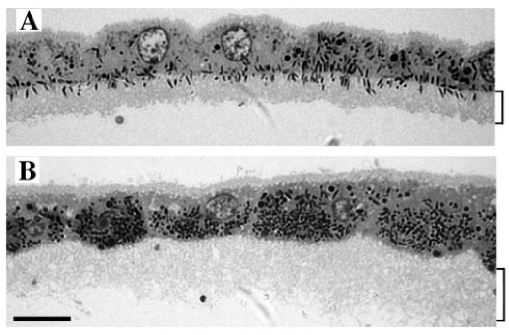
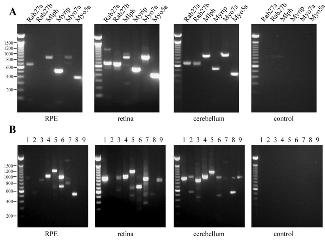
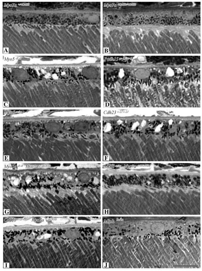
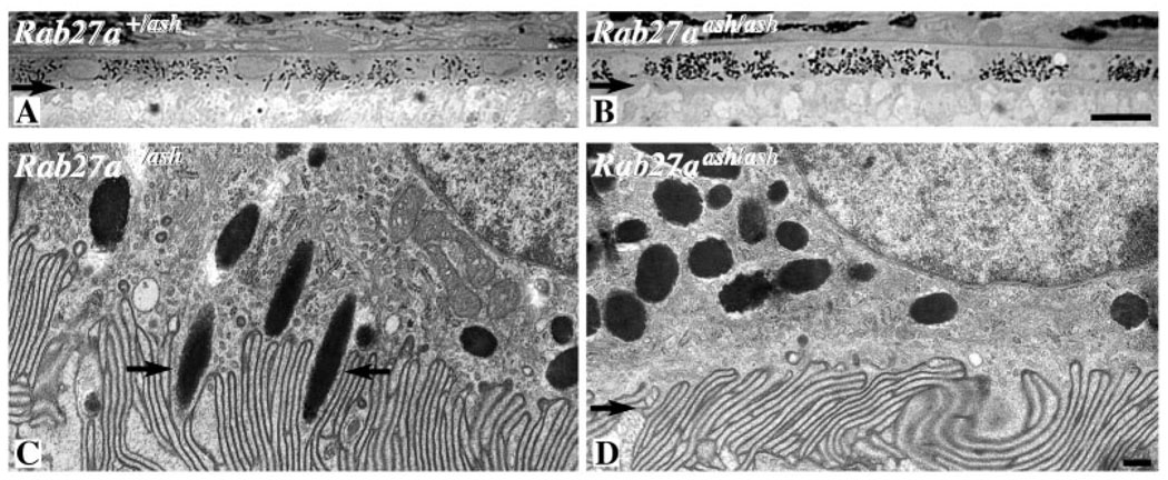
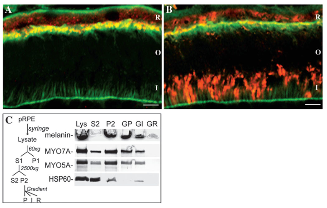
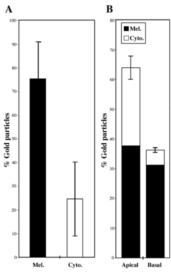
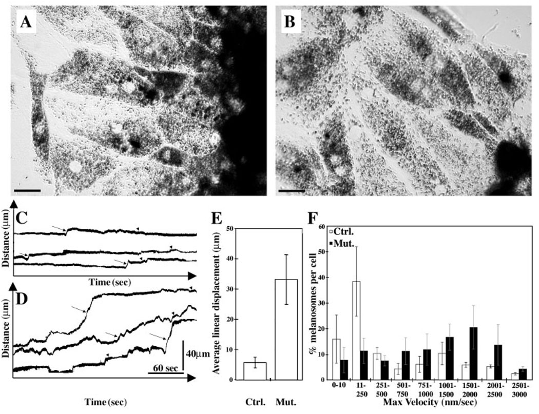
Myosin VIIa functions in the outer retina, and loss of this function causes human blindness in Usher syndrome type 1B (USH1B). In mice with mutant Myo7a, melanosomes in the retinal pigmented epithelium (RPE) are distributed abnormally. In this investigation we detected many proteins in RPE cells that could potentially participate in melanosome transport, but of those tested, only myosin VIIa and Rab27a were found to be required for normal distribution. Two other expressed proteins, melanophilin and myosin Va, both of which are required for normal melanosome distribution in melanocytes, were not required in RPE, despite the association of myosin Va with the RPE melanosome fraction. Both myosin VIIa and myosin Va were immunodetected broadly in sections of the RPE, overlapping with a region of apical filamentous actin. Some 70-80% of the myosin VIIa in RPE cells was detected on melanosome membranes by both subcellular fractionation of RPE cells and quantitative immunoelectron microscopy, consistent with a role for myosin VIIa in melanosome motility. Time-lapse microscopy of melanosomes in primary cultures of mouse RPE cells demonstrated that the melanosomes move in a saltatory manner, interrupting slow movements with short bursts of rapid movement (>1 RR01183m/second). In RPE cells from Myo7a-null mice, both the slow and rapid movements still occurred, except that more melanosomes underwent rapid movements, and each movement extended approximately five times longer (and further). Hence, our studies demonstrate the presence of many potential effectors of melanosome motility and localization in the RPE, with a specific requirement for Rab27a and myosin VIIa, which function by transporting and constraining melanosomes within a region of filamentous actin. The presence of two distinct melanosome velocities in both control and Myo7a-null RPE cells suggests the involvement of at least two motors other than myosin VIIa in melanosome motility, most probably, a microtubule motor and myosin Va.


Figures

Fig. 1
Fig. 1
Light micrographs of purified sheets of RPE cells from (A) shaker1 control (Myo7a+/4626SB) and (B) shaker1 mutant (Myo7a4626SB/4626SB) mouse retinas. Apical processes are evident as lightly stained region below the cell bodies (indicated by square brackets). In the control RPE, melanosomes are present in the apical processes, whereas the apical processes of the mutant RPE are devoid of melanosomes. Bar, 10 µm.
Fig. 2
Fig. 2
RT-PCR of purified mouse RPE cells. (A) Representative RT-PCR of mRNA expression of Rab27a, Rab27b, Melanophilin (Mlph), Myrip, Myo7a and Myo5a in purified mouse RPE cells, retina and cerebellum. Control, PCR reaction without added DNA. The position of DNA size-standards in bp is given at the left. (B) Representative RT-PCR of mRNA expression of different exophilins in purified mouse RPE cells, retina and cerebellum. 1, Exophilin 1-Rabphilin 3a; 2, Exophilin2-Granuphilin-Sytl4; 3, Exophilin3-Melanophilin-Slac2-a; 4, Exophilin4-Sytl2; 5, Exophilin5-Slac2-b, 6, Exophilin6-Sytl3; 7, Exophilin7-Sytl1; 8, Exophilin8-Myrip-Slac2-c; 9, Exophilin9-Sylt5. Control, PCR reaction without added DNA. The position of DNA size-standards in bp is given at the left.
Fig. 3
Fig. 3
Light micrographs of semithin sections of the RPE and outer segments from mice that were (A) shaker1 control (Myo7a+/4626SB), (B) shaker1 mutant (Myo7a4626SB/4626SB), (C) dilute lethal (Myo5ad-l/d-l), (D) Ames’ waltzer (Pcdh15av-3J/av-3J), (E) Snell’s waltzer (Myo6sv/sv), (F) waltzer (Cdh23v-2J/v-2J), (G) dilute lethal (Myo5ad-l/d-l) plus shaker1 heterozygote (Myo7a+/4626SB), (H) Snell’s waltzer (Myo6sv/sv) plus shaker1 heterozygote (Myo7a+/4626SB) and (I) waltzer (Cdh23v-2J/v-2J) plus shaker1 heterozygote (Myo7a+/4626SB). Genotypes are indicated on each panel. All images are the same magnification (bar, 10 µm).
Fig. 4
Fig. 4
Light (A,B) and electron (C,D) micrographs of the RPE from ashen mice. (A,C) Control (Rab27a+/ash), (B,D) mutant (Rab27aash/ash). The mice were on the C3H/HeSn background, which includes the rd1 mutation. Retinas were obtained from 14-day-old animals. No photoreceptor outer segments are evident owing to the initiation of photoreceptor degeneration, however, at this stage, the RPE remains unaffected by the degeneration. Arrows in A and B indicate the apical region of the RPE, which is completely devoid of melanosomes in the mutant. Arrows in C indicate melanosomes partially contained within the apical processes. Arrow in D indicates the apical processes, with melanosomes some distance away. A is the same magnification as B (bar, 10 µm) and C is the same as D (bar, 100 nm).
Fig. 5
Fig. 5
Mouse retinal cryosections immunolabeled with antibodies against (A) myosin VIIa or (B) myosin Va (both red), and also labeled with BODIPY-FL-phallacidin (green) to indicate actin filaments. A region of overlap is evident as a yellow band. Myosin Va is also evident in the photoreceptor inner segments. R, RPE; O, photoreceptor outer segments; I, photoreceptor inner segments. Bars, 10 µm. (C) RPE subcellular fractionation and immunoblot analysis. Left: Schematic of the fractionation procedure for purified pig RPE cells. Right: Analysis of RPE fractions. Each fraction was obtained from the same amount of starting material. Top strip: unstained stacking gel showing melanin content of each fraction, as an indication of the distribution of melanosomes. Lower strips: immunoblot labeled with myosin VIIa, myosin Va, and Hsp60 antibodies. All strips are from the same gel. Melanosomes are enriched in P2 and then in GP (and to a lesser extent in GI), following gradient separation. (GP, fraction from bottom of gradient; GI, fraction from interface; R, remainder.) Nuclei, as indicated by Trypan Blue staining were absent from GP. Labeling with Hsp60 antibody (a mitochondrial marker) indicates that mitochondria were depleted from P2 and GP.
Fig. 6
Fig. 6
Electron micrograph of the RPE from a shaker1 control (Myo7a+/4626SB) mouse retina immunolabeled with myosin VIIa antibody (10 nm gold particles). The apical region is lower right and the basal region is upper left. In the apical region of the RPE cell, melanosomes are oriented along the axis of the apical processes. Myosin VIIa is present both on the melanosome membrane (circles) and unassociated with the melanosomes, elsewhere in the cytoplasm (arrows). Insert is a magnification of the square in the picture, showing two gold particles associated with the melanosome membrane. Bar, 300 nm.
Fig. 7
Fig. 7
(A) Graph showing the percentages of gold particles, representing myosin VIIa labeling, distributed on the melanosome membrane versus those unassociated with the melanosomes but present elsewhere in the RPE cytoplasm. (B) Comparison of the labeling in the regions apical and basal to the adherens junctions of the RPE (relative amount of label on the melanosome membrane is also indicated). Data were obtained from sections with negligible background labeling (there was no labeling on the adjacent ROSs or extracellular space) and negligible labeling on simultaneously-processed, negative control sections.
Fig. 8
Fig. 8
Time-lapse microscopy of melanosome movements in cultured RPE cells. Phase-contrast micrographs of RPE cells cultured from (A) shaker1 control (Myo7a+/4626SB) and (B) shaker1 mutant (Myo7a4626SB/4626SB) mice. Bars, 20 µm. Representative kymograph traces illustrating the movements of individual melanosomes in control (C) and mutant (D) RPE cells. Arrows indicate bursts of rapid movement, and arrowheads indicate periods of slower movement. (E) Histogram illustrating the mean linear displacement resulting from single continuous rapid movements in control and mutant RPE cells (mean±s.d.). (F) Histogram illustrating the percentage of melanosomes per cell moving at different maximal velocities during 350-second periods in control and mutant RPE cells (mean±s.d.).

References

    1. Ahmed ZM, Morell RJ, Riazuddin S, Gropman A, Shaukat S, Ahmad MM, Mohiddin SA, Fananapazir L, Caruso RC, Husnain T, et al. Mutations of MYO6 are associated with recessive deafness, DFNB37. Am. J. Hum. Genet. 2003;72:1315–1322. - PMC - PubMed
    1. Astuto LM, Weston MD, Carney CA, Hoover DM, Cremers C, Wagenaar M, Moller C, Smith RJH, Pieke-Dahl S, Greenberg J, et al. Genetic heterogeneity of Usher syndrome: analysis of 151 families with Usher type I. Am. J. Hum. Genet. 2000;67:1569–1574. - PMC - PubMed
    1. Avraham KB, Hasson T, Steel KP, Kingsley DM, Russell LB, Mooseker MS, Copeland NG, Jenkins NA. The mouse Snell’s waltzer deafness gene encodes an unconventional myosin required for structural integrity of inner ear hair cells. Nat. Genet. 1995;11:369–375. - PubMed
    1. Back I, Donner KO, Reuter T. The screening effect of the pigment epithelium on the retinal rods in the frog. Vision Res. 1965;5:101–111. - PubMed
    1. Boeda B, El-Amraoui A, Bahloul A, Goodyear R, Daviet L, Blanchard S, Perfettini I, Fath KR, Shorte S, Reiners J, et al. Myosin VIIa, harmonin and cadherin 23, three Usher I gene products that cooperate to shape the sensory hair cell bundle. EMBO J. 2002;21:6689–6699. - PMC - PubMed

Publication types

MeSH terms