. 2004 Dec 7;101(49):17168-73.
doi: 10.1073/pnas.0407856101. Epub 2004 Nov 30.

Structural basis for peptidoglycan binding by peptidoglycan recognition proteins

Affiliations

Structural basis for peptidoglycan binding by peptidoglycan recognition proteins

Rongjin Guan et al. Proc Natl Acad Sci U S A. .

Abstract

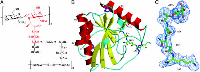
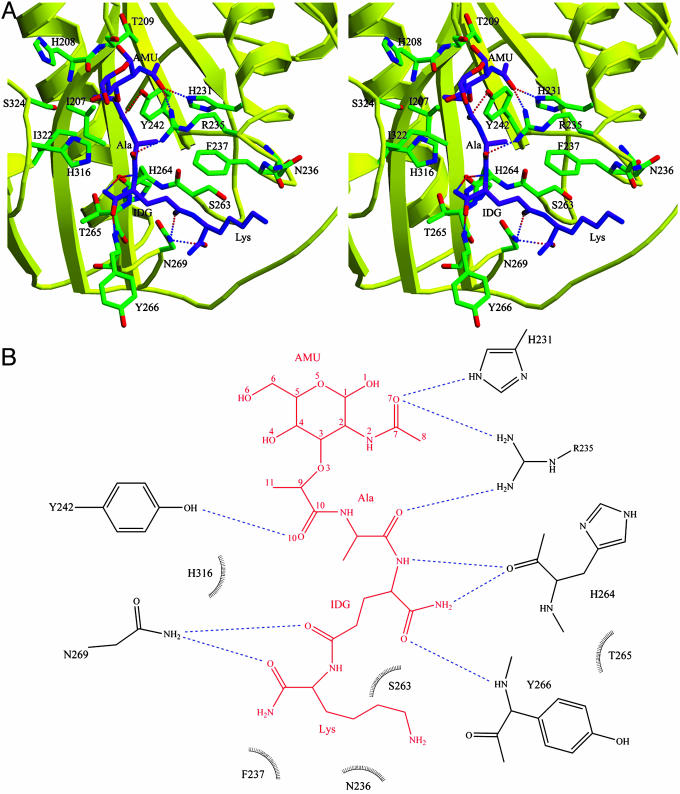
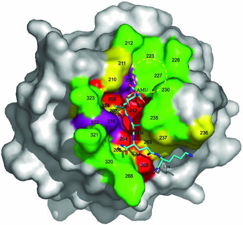
Peptidoglycan (PGN) recognition proteins (PGRPs) are pattern-recognition receptors of the innate immune system that bind and, in some cases, hydrolyze bacterial PGNs. We determined the crystal structure, at 2.30-A resolution, of the C-terminal PGN-binding domain of human PGRP-Ialpha in complex with a muramyl tripeptide representing the core of lysine-type PGNs from Gram-positive bacteria. The peptide stem of the ligand is buried at the deep end of a long binding groove, with N-acetylmuramic acid situated in the middle of the groove, whose shallow end can accommodate a linked N-acetylglucosamine. Although most interactions are with the peptide, the glycan moiety also seems to be essential for specific recognition by PGRPs. Conservation of key PGN-contacting residues shows that all PGRPs employ this basic PGN-binding mode. The structure pinpoints variable residues that likely mediate discrimination between lysine- and diaminopimelic acid-type PGNs. We also propose a mechanism for PGN hydrolysis by Zn(2+)-containing PGRPs.


Figures

Fig. 1.
Fig. 1.
Structure of Lys-type PGNs and of the PGRP-Iα–MTP complex. (A) The PGN fragment, highlighted in red, corresponds to the MTP ligand used to form the PGRP-Iα–MTP complex. Lys-type PGN peptides are usually crosslinked through a peptide bridge composed of 1–5 glycines. In parentheses is a d-alanine residue at peptide position 5 missing in PGNs from many bacteria. In Dap-type PGNs, l-lysine is replaced by meso-diaminopimelic acid, and the peptide stems are directly connected. (B) Structure of the PGRP-Iα–MTP complex. Helices are shown in red, strands in yellow, and coils in cyan. Disulfide bonds are shown in purple. The labeling of secondary structure elements follows the numbering for unbound PGRP-Iα in ref. . The N- and C-termini are indicated. The bound MTP is shown in ball-and-stick representation, with carbon atoms in green, nitrogen atoms in blue, and oxygen atoms in red. (C) σA-weighted FoFc electron density map for the MTP ligand. The contour level is 2σ. NHAc, acetamide; AMU, MurNAc; Ala, l-alanine; IDG, d-isoglutamine; Lys, l-lysine.
Fig. 2.
Fig. 2.
SPR sensograms depicting the binding of MTP versus MDP to PGRP-IαC. Concentrations of 100, 200, 300, 500, and 800 μM MTP (Left) or MDP (Right) in PBS were injected over 14,000 resonance units (RU) of immobilized PGRP-IαC at a flow rate of 20 μl/min for 180 sec. Dissociation was achieved by passing the same buffer for 300 sec.
Fig. 3.
Fig. 3.
Intermolecular contacts in the PGRP-IαC–MTP complex. (A) Stereoview of interactions between PGRP-IαC and MTP at the PGN-binding site. MTP is shown in purple, PGRP-IαC in yellow, and contacting residues in green. Hydrogen bonds are shown as dashed lines; residues forming van der Waals contacts with MTP are also highlighted. (B) Schematic representation of interactions between MTP and PGRP-IαC. MTP is shown in red; hydrogen bonds are shown as blue dashed lines. Residues making van der Waals contacts with MTP are indicated by arcs with spokes radiating toward the ligand moieties they contact. Only residues making two or more such contacts are shown. No water-mediated interactions were observed. AMU, MurNAc; IDG, d-isoglutamine.
Fig. 4.
Fig. 4.
Surface analysis of the PGN-binding site of PGRP-Iα. The molecular surface is colored according to the percentage identities of residues lining the PGN-binding groove of PGRPs based on the sequence alignments in Fig. 6. Red, >80%; purple, 60–80%; yellow, 40–60%; and green, <40%. The bound MTP is shown in ball-and-stick representation, with carbon atoms in light blue, nitrogen atoms in dark blue, and oxygen atoms in red. A putative binding pocket for the GlcNAc moiety of natural PGNs, not present in the MTP fragment, is circled in yellow. Five of 16 MTP-contacting residues in the PGRP-IαC–MTP complex, at positions 208, 231, 242, 264, and 269, are highly conserved among PGRPs (>80% identity); only two contacting residues, at positions 235 and 266, are <40% conserved. AMU, MurNAc; IDG, d-isoglutamine.

References

    1. Medzhitov, R. & Janeway, C. A., Jr. (2002) Science 296, 298–300. - PubMed
    1. Hoffmann, J. A. (2003) Nature 426, 33–38. - PubMed
    1. Weis, W. I., Taylor, M. E. & Drickamer, K. (1998) Immunol. Rev. 163, 19–34. - PubMed
    1. van Heijenoort, J. (2001) Glycobiology 11, 25R–36R. - PubMed
    1. Doyle, R. J. & Dziarski, R. (2001) in Molecular Medical Microbiology, ed. Sussman, M. (Academic, London), pp. 137–153.

Publication types

MeSH terms

Associated data

LinkOut - more resources