. 2004 Dec;24(24):10757-65.
doi: 10.1128/MCB.24.24.10757-10765.2004.

Restoration of NF-kappaB activation by tumor necrosis factor alpha receptor complex-targeted MEKK3 in receptor-interacting protein-deficient cells

Affiliations

Restoration of NF-kappaB activation by tumor necrosis factor alpha receptor complex-targeted MEKK3 in receptor-interacting protein-deficient cells

Marzenna Blonska et al. Mol Cell Biol. 2004 Dec.

Abstract

Receptor-interacting protein (RIP) plays a critical role in tumor necrosis factor alpha (TNF-alpha)-induced NF-kappaB activation. However, the mechanism by which RIP mediates TNF-alpha-induced signal transduction is not fully understood. In this study, we reconstituted RIP-deficient Jurkat T cells with a fusion protein composed of full-length MEKK3 and the death domain of RIP (MEKK3-DD). In these cells, MEKK3-DD substitutes for RIP and directly associates with TRADD in TNF receptor complexes following TNF-alpha stimulation. We found that TNF-alpha-induced NF-kappaB activation was fully restored by MEKK3-DD in these cells. In contrast, expression of a fusion protein composed of NEMO, a component of the IkappaB kinase complex, and the death domain of RIP (NEMO-DD) cannot restore TNF-alpha-induced NF-kappaB activation in RIP-deficient cells. These results indicate that the role of RIP is to specifically recruit MEKK3 to the TNF-alpha receptor complex, whereas the forced recruitment of NEMO to the TNF-alpha receptor complex is insufficient for TNF-alpha-induced NF-kappaB activation. Although MEKK2 has a high degree of homology with MEKK3, MEKK2-DD, unlike MEKK3-DD, also fails to restore TNF-alpha-induced NF-kappaB activation in RIP-deficient cells, indicating that RIP-dependent recruitment of MEKK3 plays a specific role in TNF-alpha signaling.


Figures

FIG. 1.
FIG. 1.
MEKK3 physically associates with RIP. (A) HEK293 cells (7 × 10⁵) were transiently transfected with 1 μg of Flag-tagged MEKK3 and cotransfected with 1.5 μg of Myc-tagged RIP. The cells were lysed 24 h later, and the lysates were precipitated with anti-Flag M2-agarose affinity gel. Immunocomplexes (top and middle) and whole-cell lysates (bottom) were subjected to SDS-PAGE and analyzed by Western blotting (WB) with the indicated antibodies. IP, immunoprecipitation. (B) Schematic diagram of MEKK3-DD, MEKK2-DD, and NEMO-DD fusion proteins. The RIP DD was fused to full-length MEKK3, MEKK2, or NEMO. RD, regulatory domain.
FIG. 2.
FIG. 2.
MEKK3-DD, but not the kinase-deficient mutant version of it, restores TNF-α-induced NF-κB activation in RIP-deficient cells. (A) Levels of MEKK3-DD and MEKK3(K391A)-DD fusion protein expression. RIP-deficient Jurkat cells (2 × 10⁷) stably transfected with Flag-tagged MEKK3-DD (M3-DD cell line) or MEKK3(K391A)-DD[M3(K391A)-DD cell line] were lysed and immunoprecipitated (IP) with anti-Flag M2-agarose affinity gel. Immunoprecipitates (top) and whole-cell lysates (middle and bottom) were subjected to SDS-PAGE and probed with anti-Flag antibodies or anti-RIP and anti-β-tubulin antibodies, respectively. WB, Western blotting. (B) Levels of endogenous MEKK3 in Jurkat, M3-DD, and M3(K391A)-DD cells. The cells were either untreated or stimulated with TNF-α (20 ng/ml) for 5 min. Lysates were subjected to SDS-PAGE and probed with anti-MEKK3 or anti-β-tubulin antibodies. (C) NF-κB binding activity was analyzed by EMSA. Jurkat, RIP-deficient [RIP(−)], M3-DD, and M3(K391A)-DD cells were either untreated or stimulated with TNF-α (10 ng/ml) or phorbol myristate acetate (PMA; 10 ng/ml) plus a CD28 monoclonal antibody (CD28Ab; 1 μg/ml) for 30 min. Nuclear extracts (10 μg) from these cells were incubated with 32P-labeled probes containing NF-κB binding sites for 15 min and then subjected to electrophoresis in 5% polyacrylamide gel and autoradiography. ns, nonspecific. (D) Time course of IκBα degradation in TNF-α (10 ng/ml)-stimulated cells. The level of IκBα expression was determined by Western blotting with anti-IκBα antibodies. The membrane was reprobed with anti-β-tubulin antibodies (a loading control).
FIG. 3.
FIG. 3.
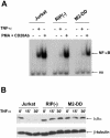
The MEKK2-DD fusion protein does not rescue TNF-α-mediated NF-κB activation in RIP-deficient cells. (A) Jurkat and RIP cells and cells expressing MEKK2-DD (M2-DD) were stimulated as described for Fig. 2C, and NF-κB binding activity was analyzed by EMSA. ns, nonspecific. (B) The expression of IκBα in TNF-α (10 ng/ml)-stimulated cells was determined by Western blotting with anti-IκBα antibodies. The membrane was reprobed with anti-β-tubulin antibodies (a loading control).
FIG. 4.
FIG. 4.
Differential effects of MEKK2-DD and MEKK3-DD expression on TNF-α-induced NF-κB activation in RIP-deficient cells. (A) Jurkat, RIP, M2-DD, and M3-DD cells were stimulated with TNF-α (10 ng/ml) for the indicated times, and EMSA was performed using 32P-labeled probes containing NF-κB or Oct-1 binding sites. ns, nonspecific. (B) Levels of MEKK2-DD and MEKK3-DD fusion protein expression. Cells (2 × 10⁷) were lysed and immunoprecipitated (IP) with anti-Flag M2-agarose affinity gel. Immunoprecipitates (top) and whole-cell lysates (bottom) were subjected to SDS-PAGE and probed with anti-Flag, anti-RIP, or anti-β-tubulin antibodies. WB, Western blotting.
FIG. 5.
FIG. 5.
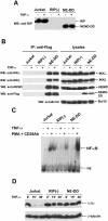
The NEMO-DD fusion protein is incorporated into the IKK complex, but its recruitment to the TNF-R1 complex is not sufficient for NF-κB activation in the absence of RIP. (A) Jurkat, RIP, and RIP cells expressing NEMO-DD (NE-DD) were either untreated or stimulated with TNF-α (10 ng/ml). The cells were lysed and subjected to SDS-PAGE and Western blotting (WB) with anti-RIP antibodies that recognize the DD of RIP. (B) Jurkat, RIP, and NE-DD cells were either untreated or stimulated with TNF-α (20 ng/ml) for 5 min. The cells were lysed and precipitated with anti-Flag M2-agarose affinity gel. Immunocomplexes (left) and whole-cell lysates (right) were subjected to SDS-PAGE and analyzed by Western blotting with antibodies against IKKα, IKKβ, NEMO, Flag, or Bcl10 as a loading control. IP, immunoprecipitation. (C) Jurkat, RIP, and NE-DD cells were stimulated as described for Fig. 2C, and NF-κB binding activity was analyzed by EMSA. PMA, phorbol myristate acetate; ns, nonspecific. (D) The expression of IκBα in TNF-α (10 ng/ml)-stimulated cells was determined by Western blotting with anti-IκBα antibodies. The membrane was reprobed with anti-β-tubulin antibodies (a loading control).
FIG. 6.
FIG. 6.
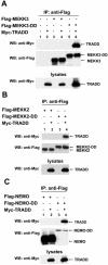
MEKK3-DD, MEKK2-DD, and NEMO-DD fusion proteins associate with TRADD. HEK293 cells (7 × 10⁵) were transiently transfected with 1.3 μg of expression plasmids encoding several constructs: Myc-tagged TRADD, Flag-tagged MEKK3, or MEKK3-DD (A), MEKK2 or MEKK2-DD (B), or NEMO or NEMO-DD (C) or with control vectors pRK6 and pcDNA3. Cells were lysed and precipitated with anti-Flag M2-agarose affinity gel. The immunocomplexes were subjected to SDS-PAGE and analyzed by Western blotting (WB) with anti-Myc or anti-Flag antibodies. The levels of expression of TRADD were assessed with anti-Myc immunoblotting of total cell lysates. IP, immunoprecipitation.
FIG. 7.
FIG. 7.
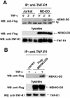
NEMO-DD and MEKK3-DD fusion proteins are recruited to the TNF-R1 complex in TNF-α-treated cells. (A) Jurkat or NE-DD cells (2 × 10⁷) were either untreated or stimulated with TNF-α (20 ng/ml) for the indicated times. These cells were lysed and precipitated with mouse monoclonal anti-TNF-R1 antibodies. The immunocomplexes (top) and whole-cell lysates (middle and bottom) were subjected to SDS-PAGE and analyzed by Western blotting (WB) with anti-Flag or anti-TNF-R1 antibodies. IP, immunoprecipitation; Ig, immunoglobulin. (B) HEK293 cells (7 × 10⁵) were transiently transfected with 1 μg of empty vector (pcDNA3) or Flag-tagged MEKK3-DD. Twenty-four hours later the cells were stimulated with TNF-α (20 ng/ml) for 10 min and lysates were tested as described for panel A.
FIG. 8.
FIG. 8.
MEKK3 and MEKK3-DD associate with IKKβ. (A) HEK293 cells (7 × 10⁵) were transiently transfected with 1.3 μg of empty vector (pcDNA3) (lane 1) or expression plasmids encoding Myc-tagged MEKK3 (lanes 2 to 4), MEKK3(K391A) (lanes 5 to 7), MEKK2 (lanes 8 to 10), or MEKK2(K395M) (lanes 11 to 13) and cotransfected with 1 μg of Flag-tagged IKKβ (lanes 3, 6, 9, and 12) or NEMO (lanes 4, 7, 10, and 13). The cells were lysed and precipitated with anti-Flag M2-agarose affinity gel. The immunocomplexes were subjected to SDS-PAGE and analyzed by Western blotting (WB) withanti-Myc antibodies. The levels of expression of the transfected products were assessed with anti-Myc and anti-Flag immunoblots of total cell lysates. IP, immunoprecipitation. (B) HEK293 cells (7 × 10⁵) were transiently transfected with 1.3 μg of Myc-tagged MEKK3 (lanes 2, 5, and 6) and cotransfected with Flag-tagged IKKβ or NEMO. The cells were lysed and tested as described for panel A. (C) HEK293 cells (7 × 10⁵) were transiently transfected with Flag-tagged MEKK3, MEKK3-DD, NEMO, or NEMO-DD expression vectors, and the cells were lysed 24 h later. The lysates were precipitated with anti-Flag M2-agarose affinity gel. Immunocomplexes (top and middle) and whole-cell lysates (bottom) were subjected to SDS-PAGE and analyzed by Western blotting with antibodies against IKKβ or Flag.
FIG. 9.
FIG. 9.
Kinase-active form of MEKK3 phosphorylates IKKβ. HEK293 cells (7 × 10⁵) were transiently transfected with 1.3 μg of empty vector (pcDNA3) (lane 1) or expression plasmids encoding Myc-MEKK3 (lane 2) and Myc-MEKK3(K391A) (lane 3). The cells were lysed 24 h later and precipitated with anti-c-Myc-agarose affinity gel. The immunocomplexes were subjected to an in vitro kinase assay. Whole-cell lysates were analyzed by Western blotting (WB) with anti-phospho-IKKβ and anti-IKKβ antibodies. IP, immunoprecipitation.
FIG. 10.
FIG. 10.
Working model of TNF-α-induced NF-κB activation in RIP-deficient cells expressing MEKK3-DD or NEMO-DD fusion proteins.

References

    1. Blank, J. L., P. Gerwins, E. M. Elliott, S. Sather, and G. L. Johnson. 1996. Molecular cloning of mitogen-activated protein/ERK kinase kinases (MEKK) 2 and 3. Regulation of sequential phosphorylation pathways involving mitogen-activated protein kinase and c-Jun kinase. J. Biol. Chem. 271:5361-5368. - PubMed
    1. Bouwmeester, T., A. Bauch, H. Ruffner, P. O. Angrand, G. Bergamini, K. Croughton, C. Cruciat, D. Eberhard, J. Gagneur, S. Ghidelli, C. Hopf, B. Huhse, R. Mangano, A. M. Michon, M. Schirle, J. Schlegl, M. Schwab, M. A. Stein, A. Bauer, G. Casari, G. Drewes, A. C. Gavin, D. B. Jackson, G. Joberty, G. Neubauer, J. Rick, B. Kuster, and G. Superti-Furga. 2004. A physical and functional map of the human TNF-alpha/NF-kappa B signal transduction pathway. Nat. Cell Biol. 6:97-105. - PubMed
    1. Chen, G., and D. V. Goeddel. 2002. TNF-R1 signaling: a beautiful pathway. Science 296:1634-1635. - PubMed
    1. Devin, A., A. Cook, Y. Lin, Y. Rodriguez, M. Kelliher, and Z. Liu. 2000. The distinct roles of TRAF2 and RIP in IKK activation by TNF-R1: TRAF2 recruits IKK to TNF-R1 while RIP mediates IKK activation. Immunity 12:419-429. - PubMed
    1. Ellinger-Ziegelbauer, H., K. Brown, K. Kelly, and U. Siebenlist. 1997. Direct activation of the stress-activated protein kinase (SAPK) and extracellular signal-regulated protein kinase (ERK) pathways by an inducible mitogen-activated protein kinase/ERK kinase kinase 3 (MEKK) derivative. J. Biol. Chem. 272:2668-2674. - PubMed

Publication types

MeSH terms

Substances