. 2005 May 1;387(Pt 3):849-58.
doi: 10.1042/BJ20041836.

Identification of a unique filamin A binding region within the cytoplasmic domain of glycoprotein Ibalpha

Affiliations

Identification of a unique filamin A binding region within the cytoplasmic domain of glycoprotein Ibalpha

Susan L Cranmer et al. Biochem J. .

Abstract

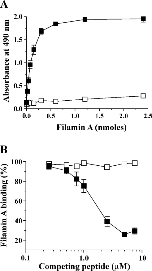
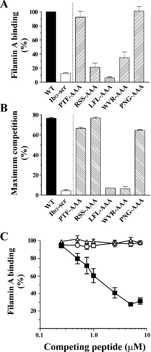
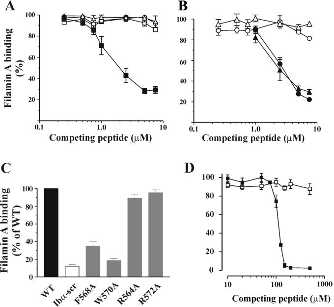
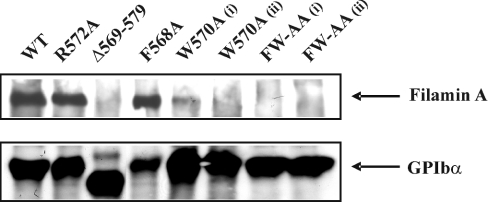
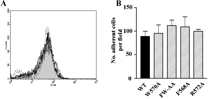
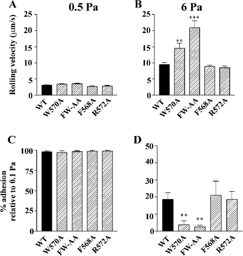
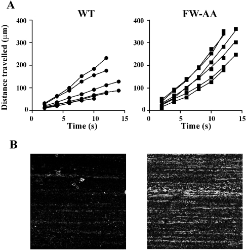
Binding of the platelet GPIb/V/IX (glycoprotein Ib/V/IX) receptor to von Willebrand factor is critical for platelet adhesion and aggregation under conditions of rapid blood flow. The adhesive function of GPIbalpha is regulated by its anchorage to the membrane skeleton through a specific interaction with filamin A. In the present study, we examined the amino acid residues within the cytoplasmic tail of GPIbalpha, which are critical for association with filamin A, using a series of 25-mer synthetic peptides that mimic the cytoplasmic tail sequences of wild-type and mutant forms of GPIbalpha. Peptide binding studies of purified human filamin A have demonstrated a major role for the conserved hydrophobic stretch L567FLWV571 in mediating this interaction. Progressive alanine substitutions of triple, double and single amino acid residues within the Pro561-Arg572 region suggested an important role for Trp570 and Phe568 in promoting GPIbalpha binding to filamin A. The importance of these two residues in promoting filamin A binding to GPIbalpha in vivo was confirmed from the study of Chinese-hamster ovary cells expressing GPIbalpha Trp570-->Ala and Phe568-->Ala substitutions. Phenotypic analysis of these cell lines in flow-based adhesion studies revealed a critical role for these residues in maintaining receptor anchorage to the membrane skeleton and in maintaining cell adhesion to a von Willebrand factor matrix under high-shear conditions. These studies demonstrate a novel filamin A binding motif in the cytoplasmic tail of GPIbalpha, which is critically dependent on both Trp570 and Phe568.


Figures

Figure 1
Figure 1. Binding of filamin A to WT and scrambled GPIbα peptides
(A) To define the experimental conditions required to quantify the direct binding capacity of filamin A to WT and scrambled GPIbα peptides, filamin A (0–2.4 nmol) was incubated with 15 pmol of either immobilized biotinylated WT (■) or scrambled (Ibα-scr, □) peptide for 60 min as described in the Materials and methods section. Bound filamin A was detected using NCL-FIL mAb and HRP-conjugated secondary reagents for colorimetric detection at 490 nm as described in the Materials and methods section. Absorbances are presented as the means±S.D. for two experiments. Filamin A binding to WT peptide was saturable and reached a maximum between 0.6 and 1.2 nmol. (B) In competition binding experiments, 0.01–1 nmol of either soluble WT (■) or scrambled (Ibα-scr, □) peptide was incubated with filamin A (1.2 nmol) for 60 min, then incubated for a further 60 min with immobilized WT peptide. The level of bound filamin A was measured as described above and expressed relative to the absorbance detected for filamin A binding to immobilized WT peptide in the absence of competing soluble peptide, which was assigned as 100%. The results are presented as the means±S.E.M. for three experiments.
Figure 2
Figure 2. Amino acid sequences of wild-type, scrambled and alanine-substituted GPIbα peptides used in the present study
Figure 3
Figure 3. Binding of filamin A to triple alanine-substituted GPIbα peptides
(A) Direct binding of filamin A to triple alanine-substituted peptides was performed by incubating 1.2 nmol filamin A with 15 pmol immobilized peptide as described in the Materials and methods section. Results (means±S.E.M. for four experiments) are expressed as the percentage of filamin A binding, relative to the binding measured for immobilized WT peptide which was assigned as 100%. (B) Competition binding experiments were performed as described in Figure 1 and the results (means±S.E.M. for three experiments) are expressed as the maximum percentage of competition. (C) Competition binding curves are shown for WT (■), LFL-AAA (△) and WVR-AAA (○). The level of bound filamin A is expressed relative to the binding detected for filamin A binding to immobilized WT peptide in the absence of competing soluble peptide, which was assigned as 100%. The data presented are the means±S.E.M. for six experiments.
Figure 4
Figure 4. Binding of filamin A to double and single alanine-substituted peptides and 5-mer peptides
(A) Competition binding experiments using double alanine-substituted peptides were performed as described for Figure 1. Competition binding curves are shown for WT (■), FW-AA (△), LF-AA (○) and FL-AA (□) and results are expressed as the percentage of filamin A binding relative to the binding measured for immobilized WT peptide in the absence of competing peptide, assigned as 100%. The results presented are the means±S.E.M. for four experiments and demonstrated that none of the double alanine mutants were able to compete with WT for filamin A binding. (B) Competition binding curves for single alanine mutants, F568A (△), W570A (○), R564A (▲) and R572A (●). The results presented are the means±S.E.M. for four experiments. Neither F568A nor W570A demonstrated any competition with WT for filamin A binding, whereas R564A and R572A exhibited similar competition as WT peptide. (C) Direct binding of filamin A to single alanine-substituted peptides was performed as described in the Materials and methods section. Filamin A binding to substituted peptides (means±S.E.M. for three or four experiments) is expressed as percentage of binding relative to WT, which was assigned as 100%. (D) Competition binding curves for 5-mer peptides LFLWV (■) and LFLAV (□). The results presented are the means±S.E.M. for five experiments.
Figure 5
Figure 5. Association of WT and mutant forms of GPIbα with filamin A
Adherent CHO cells (8×10⁶/ml) were detached, incubated with the membrane-permeable EZ-link-N-hydroxysuccinimidobiotin and lysed with 1% Triton X-100. GPIb–IX was immunoprecipitated using the anti-GPIbβ mAb (RAM.1), as described in the Materials and Methods section. Immunoprecipitated proteins were separated by SDS/PAGE (5% gel), transferred on to PVDF membranes, and the immunoprecipitated bands were detected by enhanced chemiluminescence. Mutation of Trp570 to alanine almost completely eliminated the co-immunoprecipitation of filamin A. The double mutation of Phe568 and Trp570 to alanine resulted in a complete disruption of the interaction between GPIb–IX complex and filamin A. These results are from one experiment, representative of five.
Figure 6
Figure 6. Analysis of GPIbα surface expression and the ability of CHO cells to adhere to vWf under shear conditions
(A) Surface expression of GPIbα was analysed by flow cytometry as described in the Materials and methods section. GPIbα expression was similar for all cell lines; WT (grey-filled histogram), W570A (thick grey line), FW-AA, (black dotted line), F568A (thin black line) and R572A (thick black line). (B) Cells were perfused through BvWf-coated microslides (10 μg/ml) at a shear stress of 0.1 Pa. After 5 min of perfusion, the mean number of adherent cells/field over 5 fields was calculated and the results are expressed as the number of adherent cells/field (means±S.E.M.) for four to seven experiments.
Figure 7
Figure 7. Effects of alanine substitutions on GPIb–IX-dependent CHO cell-rolling velocity and detachment under shear conditions
CHO cells were perfused through BvWf-coated microslides (10 μg/ml) for 5 min at a shear stress of 0.1 Pa, followed by stepwise increases in shear stress up to 6 Pa. (A, B) Cell-rolling velocities were analysed as described in the Materials and methods section and the results presented are the means±S.E.M. for four to seven experiments. At 0.5 Pa there were no significant differences in rolling velocities between WT and any of the mutant cell lines, whereas at 6 Pa, only W570A and FW-AA cells exhibited significantly faster velocities when compared with WT (P<0.01 and 0.001 respectively). (C, D) Cell detachment was analysed in the same experiments as described in the Materials and methods section and the results are presented as means±S.E.M. There was essentially no detachment of any of the cell lines at 0.5 Pa, but at 6 Pa both W570A and FW-AA were significantly less capable of remaining adherent (P<0.01 in both cases) when compared with WT. There was no significant difference between the adhesion of F568A and R572A cells compared with WT, for both rolling velocity and their ability to remain adherent at high shear stress.
Figure 8
Figure 8. Time-dependent changes to rolling velocity in detaching WT and FW-AA cells and extraction of GPIb–IX from the membrane under high-shear-stress conditions
(A) Rolling velocities before cell detachment were analysed for WT and FW-AA cells (six cells for each cell line) by measuring the distance travelled over 2 s intervals for the 10–12 s period immediately before their detachment. WT cells exhibited a constant rate of translocation before detachment, whereas FW-AA cells exhibited a considerable time-dependent increase in translocation rate before the point of detachment. (B) Analysis of receptor extraction was performed as described in the Materials and methods section. Two representative confocal microscopic images are shown for each cell line from one experiment, representative of four independent experiments. Images were captured using identical photomultiplier tube settings on the confocal microscope and clearly demonstrate, for FW-AA cells, the presence of GPIbα receptor tracks after cells had been detached from the vWf matrix under high-shear conditions.

Similar articles

Cited by

References

    1. Savage B., Saldivar E., Ruggeri Z. M. Initiation of platelet adhesion by arrest onto fibrinogen or translocation on von Willebrand factor. Cell (Cambridge, Mass.) 1996;84:289–297. - PubMed
    1. Berndt M. C., Shen Y., Dopheide S. M., Gardiner E. E., Andrews R. K. The vascular biology of the glycoprotein Ib-IX-V complex. Thromb. Haemostasis. 2001;86:178–188. - PubMed
    1. Lopez J. A., Andrews R. K., Afshar-Kharghan V., Berndt M. C. Bernard-Soulier syndrome. Blood. 1998;91:4397–4418. - PubMed
    1. Okita J. R., Pidard D., Newman P. J., Montgomery R. R., Kunicki T. J. On the association of glycoprotein Ib and actin-binding protein in human platelets. J. Cell Biol. 1985;100:317–321. - PMC - PubMed
    1. Du X., Fox J. E., Pei S. Identification of a binding sequence for the 14-3-3 protein within the cytoplasmic domain of the adhesion receptor, platelet glycoprotein Ib alpha. J. Biol. Chem. 1996;271:7362–7367. - PubMed

Publication types