Review
. 2005 Jan 1:10:216-35.
doi: 10.2741/1522. Print 2005 Jan 1.

Pathobiology of dynorphins in trauma and disease

Affiliations
Review

Pathobiology of dynorphins in trauma and disease

Kurt F Hauser et al. Front Biosci. .

Abstract

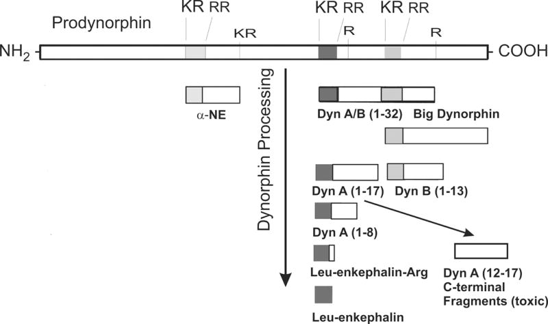
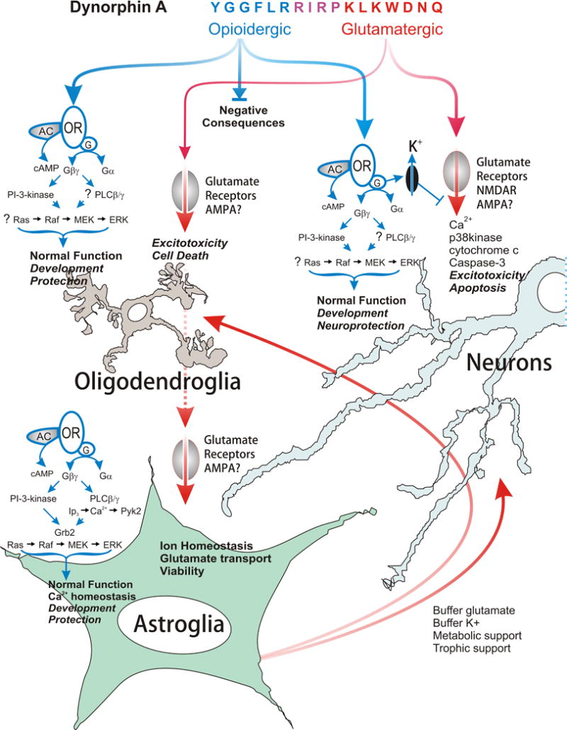
Dynorphins, endogenous opioid neuropeptides derived from the prodynorphin gene, are involved in a variety of normative physiologic functions including antinociception and neuroendocrine signaling, and may be protective to neurons and oligodendroglia via their opioid receptor-mediated effects. However, under experimental or pathophysiological conditions in which dynorphin levels are substantially elevated, these peptides are excitotoxic largely through actions at glutamate receptors. Because the excitotoxic actions of dynorphins require supraphysiological concentrations or prolonged tissue exposure, there has likely been little evolutionary pressure to ameliorate the maladaptive, non-opioid receptor mediated consequences of dynorphins. Thus, dynorphins can have protective and/or proapoptotic actions in neurons and glia, and the net effect may depend upon the distribution of receptors in a particular region and the amount of dynorphin released. Increased prodynorphin gene expression is observed in several disease states and disruptions in dynorphin processing can accompany pathophysiological situations. Aberrant processing may contribute to the net negative effects of dysregulated dynorphin production by tilting the balance towards dynorphin derivatives that are toxic to neurons and/or oligodendroglia. Evidence outlined in this review suggests that a variety of CNS pathologies alter dynorphin biogenesis. Such alterations are likely maladaptive and contribute to secondary injury and the pathogenesis of disease.


Figures

Figure 1
Figure 1
Summary of the major posttranslational products of prodynorphin. Prodynorphin is converted into multiple bioactive peptide fragments (3,224,225), including alpha-Neoendorphin (α-NE), Big Dynorphin (Dyn AB 1–32), Leumorphin (Dyn B 1–29), dynorphin A (Dyn A 1–17), dynorphin B (Dyn B 1–16), Leucine-enkephalin-arginine (Leu-enkephalin-Arg), and potentially Leucine-enkephalin (Leu-enkephalin). Processing is likely to be altered following injury or disease. Additional bioactive derivatives of prodynorphin likely exist besides those shown here. Many of the C-terminal fragments of dynorphin A, which are intrinsically neurotoxic, can be isolated from neural tissue in vivo or in vitro. Prodynorphin is highly basic, containing multiple lysine (K) and arginine (R) residues.
Figure 2
Figure 2
Summary of the neuroprotective and deleterious affects of dynorphin A (1–17) in neurons, astroglia, and oligodendroglia. Dynorphin A has intrinsic activity at both opioid and non-opioid, i.e., glutamatergic receptors. The N-terminal peptide derivatives are active at opioid receptors, whereas C-terminal-derived fragments can activate glutamate receptors. At physiological concentrations, dynorphin functions normally by activating kappa-opioid receptors (KOR). The activation of opioid receptors can have beneficial effects in isolated neurons, but may also have negative consequences depending on the particular neural cell type affected, as well as the nature and timing of the insult. By contrast, at supraphysiological levels, dynorphin A acts via glutamate receptors and has excitotoxic effects in neurons and oligodendroglia, and potentially destabilizes astroglia. Abbreviations: adenylyl cyclase (AC); alpha-amino-3-hydroxy-5-methylisoxazole-4-propionate receptors (AMPA); cyclic AMP (cAMP); extracellular signal-regulated protein kinase (ERK); growth factor receptor-bound protein 2 (Grb2); G-protein complex (G); G-protein-alpha subunit (Gα); G-protein-beta-gamma subunit complex (Gβγ); inositol trisphosphate (IP3); p38 mitogen-activated protein kinase (p38kinase); N-methyl-D-aspartate receptors (NMDAR); phosphatidylinositol 3-kinase (PI-3-kinase), phospholipase C-beta-gamma (PLCβ/γ); proline-rich tyrosine kinase-2 (Pyk2); protein kinase C (PKC);.
Figure 3
Figure 3
Primary sequence of big dynorphin, dynorphin A and dynorphin B. Big dynorphin has intrinsic biological activity that differs from both dynorphin A and dynorphin B. Big dynorphin’s novel effects are not mediated by opioid receptors. The highly basic nature of the big dynorphin peptide may permit big dynorphin to interact selectively with other target molecules (46).

References

    1. Kakidani H, Furutani Y, Takahashi H, Noda M, Morimoto Y, Hirose T, Asai M, Inayama S, Nakanishi S, Numa S. Cloning and sequence analysis of cDNA for porcine beta-neo- endorphin/dynorphin precursor. Nature. 1982;298:245–249. - PubMed
    1. Goldstein A, Tachibana S, Lowney LI, Hunkapiller M, Hood L. Dynorphin-(1–13), an extraordinarily potent opioid peptide. Proc Natl Acad Sci U S A. 1979;76:6666–6670. - PMC - PubMed
    1. Evans CJ, Hammond DL, Pasternak GW, editors. The Opiate Receptors. Humana; Clifton, New Jersey: pp. 23–71.
    1. Chavkin C, James IF, Goldstein A. Dynorphin is a specific endogenous ligand of the kappa opioid receptor. Science. 1982;215:413–415. - PubMed
    1. Goldstein A. Binding selectivity profiles for ligands of multiple receptor types: focus on opioid receptors. Trends Pharmacol Sci. 1987;8:456–459.

Publication types

LinkOut - more resources