Comparative Study
. 2004 Dec 15;18(24):3010-5.
doi: 10.1101/gad.1250804. Epub 2004 Dec 1.

A role for beta-actin in RNA polymerase III transcription

Affiliations
Comparative Study

A role for beta-actin in RNA polymerase III transcription

Ping Hu et al. Genes Dev. .

Abstract

When transcription from the human U6 snRNA gene is reconstituted with recombinant factors and purified RNA polymerase III (pol III), pol III must be treated with CK2 to be active. We show that highly purified pol III contains associated beta-actin, and beta-actin localizes to an active U6 promoter in vivo. Pol III immunoprecipitated from IMR90 cells treated with a genotoxic agent lacks associated beta-actin and is inactive in the reconstituted assay. Transcription is regained upon treatment of pol III with CK2 and addition of beta-actin. This suggests that beta-actin associated with pol III is essential for basal pol III transcription.


Figures

Figure 1.
Figure 1.
β-actin is associated with pol III. (A) Purification scheme of tagged pol III from a HeLa cell line expressing Flag- and His-tagged RPC4. (B) β-actin and RPC1 copurify during mono Q chromatography. This panel is reprinted from Figure 4D in Hu et al. (2003) (© 2003 with permission from Elsevier) and shows immunoblots performed with anti-RPC1 and anti-β-actin antibodies. (C) Purification scheme of endogenous pol III. (D) β-actin and pol III copurify during sucrose gradient fractionation. The immunoblot was probed as in B. (E) The association of β-actin with pol III is resistant to high salt. HeLa whole-cell extracts were incubated with anti-RPC4 beads, the beads were then washed with 0.6, 1, or 1.5 M KCl as indicated above the lanes, and the bound material was eluted, fractionated on a gel, and immunoblotted with an anti-β-actin antibody. In lane 4, preimmune antibody beads were used. (F) HeLa whole-cell extract was incubated with anti-RPC1 beads, the beads were washed with 0.6 M KCl, and the bound material was analyzed as in E. (G) As in F except that the HeLa whole-cell extract was incubated with anti-RPC5 beads.
Figure 4.
Figure 4.
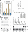
β-actin is required for U6 transcription in vitro. (A) β-actin dissociates from an active U6 promoter upon MMS treatment. The Y-axis indicates the ratio of DNA amplified from actively growing IMR90 cells to DNA amplified from MMS-treated IMR90 cells. The error bars are based on three independent PCR reactions with the same templates and primers. (B) Extracts from MMS-treated IMR90 cells show reduced pol III transcription. Extracts from actively growing (lane 1) and MMS-treated (lanes 2-5) IMR90 cells were tested for transcription from the U6 and VAI promoters. (IC) Internal control for RNA recovery. (C) Equal amounts of pol III were precipitated from untreated and IMR90-treated cells. Pol III was immunoprecipitated with an anti-RPC4 antibody, and the bound material was analyzed by immunoblotting with anti-RPC1 antibodies. (D) β-actin is required for U6 transcription in a reconstituted transcription system. SNAPc purified from baculovirus-infected insect cells was treated with 60 μM LY294002 followed by 5 U of CK2 holoenzyme (SNAPc +I). TBP, Brf2, and Bdp1 were purified from E. coli. These factors were supplemented with 6 μL of pol III immunoprecipitated from actively growing (lane 1) or 6 μL (lanes 2,4,6-9) and 12 μL (lanes 3,5) of pol III immunoprecipitated from MMS-treated IMR90 cells. In lanes 4, 5, 8, and 9, immunoprecipitated pol III was incubated with 5 U of CK2 holoenzyme for 10 min at 30°C followed by addition of 60 μM LY294002 (CK2). In lanes 6 and 7, immunoprecipitated pol III was incubated first with LY294002 and then with CK2. In lanes 6-9, increasing amounts (50 and 100 ng) of β-actin were added to the reactions. The various combinations of factors were then tested for U6 transcription.
Figure 2.
Figure 2.
β-actin can be localized to the promoter sequences of an actively transcribed U6 gene. Cross-linked and sonicated chromatin from actively growing Hela (top panel) or IMR90 (bottom panel) cells was incubated with either no antibody (lanes 4,5), or antibodies against the SNAP190 subunit of SNAPc (CS696, lanes 6,7), TFIIB (CS396, lanes 8,9), or β-actin (AC-74, Sigma, lanes 10,11). In each case, the immunoprecipitated material was analyzed by PCR with test primers (T) specific for the promoter of an actively transcribed U6 gene, and control primers (C) specific for the promoter of an inactive U6 gene. In lanes 2 and 3, the starting chromatin material was directly analyzed by PCR. The order of the lanes in the bottom panel, which are from the same gel, has been rearranged for clarity.
Figure 3.
Figure 3.
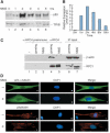
β-actin dissociates from pol III upon MMS treatment in IMR90 cells. (A) p53 peaks 2 h after MMS treatment. IMR90 cells were treated with 1 mM MMS. Samples were analyzed by immunoblotting with anti-p53 and anti-TFIIB antibodies at the times indicated after MMS treatment. (B) Quantitation of the p53 signals in A after normalization to the TFIIB internal control. (C) β-actin dissociates from pol III after MMS treatment. Extracts from either actively growing (lanes 2,4) or MMS-treated (lanes 3,5) IMR90 cells were incubated with preimmune (lanes 2,3) or anti-RPC4 (lanes 4,5) antibody beads, the beads were washed with 0.6 M KCl, and the bound material was eluted and analyzed by immunoblotting with anti-β-actin and anti-RPC1 antibodies. In lane 1, the anti-RPC4 beads were not incubated with extract prior to immunoblotting analysis of the bound material. Lanes 6 and 7 show immunoblots of the starting material that was used for the immunoprecipitations. (D) MMS treatment does not change the overall cytoskeleton structure. Actively growing (panels a-c,g-i) and MMS-treated (panels d-f,j-l) IMR90 cells were stained with DAPI and either an immunofluorescent-labeled anti-β-tubulin antibody or Alexa Fluor 568-labeled phalloidin, which stains filamentous actin.

References

    1. Ascough K.R. 2004. Endocytosis: Actin in the driving seat. Curr. Biol. 14: R124-R126. - PubMed
    1. Bartholomew B., Durkovich, D., Kassavetis, G.A., and Geiduschek, E.P. 1993. Orientation and topography of RNA polymerase III in transcription complexes. Mol. Cell. Biol. 13: 942-952. - PMC - PubMed
    1. Bettinger B.T., Gilbert, D.M., and Amberg, D.C. 2004. Actin up in the nucleus. Nat. Rev. Mol. Cell. Biol. 5: 410-415. - PubMed
    1. Brakebusch C. and Fassler, R. 2003. The integrin-actin connection, an eternal love affair. EMBO J. 22: 2324-2333. - PMC - PubMed
    1. Cramer P., Bushnell, D.A., and Kornberg, R.D. 2001. Structural basis of transcription: RNA polymerase II at 2.8 angstrom resolution. Science 292: 1863-1876. - PubMed

Publication types

LinkOut - more resources