Comparative Study
. 2005 Feb;88(2):1423-31.
doi: 10.1529/biophysj.104.052746. Epub 2004 Dec 1.

Molecular force modulation spectroscopy revealing the dynamic response of single bacteriorhodopsins

Affiliations
Comparative Study

Molecular force modulation spectroscopy revealing the dynamic response of single bacteriorhodopsins

Harald Janovjak et al. Biophys J. 2005 Feb.

Abstract

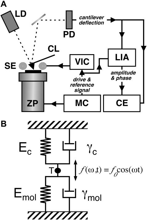
Recent advances in atomic force microscopy allowed globular and membrane proteins to be mechanically unfolded on a single-molecule level. Presented is an extension to the existing force spectroscopy experiments. While unfolding single bacteriorhodopsins from native purple membranes, small oscillation amplitudes (6-9 nm) were supplied to the vertical displacement of the cantilever at a frequency of 3 kHz. The phase and amplitude response of the cantilever-protein system was converted to reveal the elastic (conservative) and viscous (dissipative) contributions to the unfolding process. The elastic response (stiffness) of the extended parts of the protein were in the range of a few tens pN/nm and could be well described by the derivative of the wormlike chain model. Discrete events in the viscous response coincided with the unfolding of single secondary structure elements and were in the range of 1 microNs/m. In addition, these force modulation spectroscopy experiments revealed novel mechanical unfolding intermediates of bacteriorhodopsin. We found that kinks result in a loss of unfolding cooperativity in transmembrane helices. Reconstructing force-distance spectra by the integration of amplitude-distance spectra verified their position, offering a novel approach to detect intermediates during the forced unfolding of single proteins.


Figures

FIGURE 1
FIGURE 1
Illustration of the experimental setup. (A) A commercial AFM with an optical detection system (laser diode (LD) and photodetector (PD)) was equipped with magnetically coated cantilevers (CL) and a magnetic excitation system consisting of a solenoid (SE). The solenoid was driven by a voltage-current converter (VIC) connected to the sinusoidal drive signal from the microscope controller (MC). While controlling the z-position of the piezoelectric actuator (ZP), the cantilever deflection was analyzed in a lock-in amplifier (LIA) to separate amplitude and phase of the oscillation. Amplitude and phase were recorded together with the quasi-static deflection of the cantilever in external capture electronics (CE). (B) The cantilever-molecule system was considered as two VK elements (i.e., a spring and dashpot) acting in parallel, as the motion of the cantilever is detected at its tip (T).
FIGURE 2
FIGURE 2
Unfolding pathways of individual BRs. (A) Conventional F-D curve (left) showing a typical unfolding spectrum of a single BR together with the schematic unfolding pathway (right). The first peaks detected at tip-sample separations below 15 nm indicate the unfolding of helices F and G. However, nonspecific interactions between the membrane surface and AFM tip make a detailed analysis of the first peaks difficult. After unfolding these elements, 88 aa are tethered between the tip and the surface (a). Separating the tip further from the surface stretches the polypeptide (b), thereby exerting force to helices E and D. At a certain critical load, helices E and D unfold in one event. As the number of aa linking the tip and the surface is now increased to 148, the cantilever relaxes (c). In a next step, 148 aa are extended, thereby pulling on helix C (d). After unfolding helices B and C in a single step, the molecular bridge is lengthened to 219 aa (e). By further separating tip and membrane, helix A unfolds (f) and the polypeptide is completely extracted from the membrane (g). (B and C) Unfolding individual secondary structure elements. (B) Occasionally the first unfolding peak (88 aa) shows two shoulder peaks, which indicate the stepwise unfolding of the helical pair. If both shoulders occur, the peak at 88 aa indicates the unfolding of helix E, the peak at 94 aa of loop D-E, and the peak at 105 aa corresponds unfolding of helix D. (C) The shoulder peaks of the second peak indicate the stepwise unfolding of helices C and B and loop B-C. The peak at 148 aa indicates the unfolding of helix C, the peak at 158 aa of the loop BC, and the peak at 175 aa represents unfolding of helix B.
FIGURE 3
FIGURE 3
Force modulation spectroscopy of single BRs. (A) A superimposition of 15 F-D curves each recorded while unfolding a single BR molecule. The overlaid curves show a reproducible unfolding pattern similar to that observed in conventional unfolding experiments of BR (Fig. 2 A). (B and C) Application of a small oscillation to the cantilever allows the measurement of the amplitude (B) and phase (C) response of single proteins. The A-D and phase-distance curves were superimposed with the same distance offsets as the corresponding F-D curves. The amplitude of the cantilever oscillation decreased by up to ≈50% during the force curve, whereas the phase response showed less clear events.
FIGURE 4
FIGURE 4
Elastic and dissipative response of BR. The elasticity of the extended parts of BR as well as the relaxation time and the damping coefficient corresponding to unfolding events are derived from the amplitude and phase response. (A) Fitting the elasticity curves with the derivative of the WLC model (Eq. 8) indicates the characteristic unfolding spectrum of BR to consist of purely elastic polypeptide extension. (B and C) Discrete events are observed in the damping coefficient and the relaxation time of the molecules.
FIGURE 5
FIGURE 5
Force modulation spectroscopy reveals new unfolding intermediates. Additional unfolding events were observed during forced modulation unfolding of BR indicating the presence of novel unfolding intermediates. In the left frames, the curves from the conventional pulling experiment (Fig. 2, B and C) are shown in gray, whereas individual force modulation F-D curves are overlaid. The selected curves show the three new unfolding peaks, each of which was detected in ≈50% of all curves. Fitting these peaks with the WLC model revealed that they correspond to the extension of 76, 125 (A), and 195 (B) aas. As for any of the other force events, the positions of the peaks allow localizing the corresponding unfolding barriers and unfolding intermediates in the structure of the protein (right frames).
FIGURE 6
FIGURE 6
Reconstruction of a F-D curve. (A) Experimental F-D curve (shaded line) was reconstructed from the corresponding A-D curve in several segments (solid black lines flanked by two arrowheads). The x-position of the arrowheads corresponds to the point where the integration of each segment was started (solid arrowheads) or stopped (open arrowheads). Consequently, the y-positions of the solid arrowheads correspond to the constant C in Eq. 10 for each segment. Excellent agreement between reconstructed and measured data is obtained for areas of elastic polypeptide extension, whereas no agreement was observed if the reconstruction was performed over force peaks. (B) For each segment,a difference curve was calculated by subtracting the measured data from the reconstructed data. As the poor overlap between reconstructed and measured data leads to a sudden increase in the difference curve (arrowheads), this approach can be used to detect unfolding events in F-D curves. For the detection routine, we have used a threshold of 20 pN (dashed line).

Comment in

  • Unraveling bacteriorhodopsin.
    van Noort J. van Noort J. Biophys J. 2005 Feb;88(2):763-4. doi: 10.1529/biophysj.104.056242. Epub 2004 Dec 13. Biophys J. 2005. PMID: 15596486 Free PMC article. No abstract available.

References

    1. Baldwin, J. M. 1993. The probable arrangement of the helices in G protein-coupled receptors. EMBO J. 12:1693–1703. - PMC - PubMed
    1. Best, R. B., B. Li, A. Steward, V. Daggett, and J. Clarke. 2001. Can nonmechanical proteins withstand force? Stretching barnase by atomic force microscopy and molecular dynamics simulation. Biophys. J. 81:2344–2356. - PMC - PubMed
    1. Binnig, G., C. F. Quate, and C. Gerber. 1986. Atomic force microscope. Phys. Rev. Lett. 56:930–933. - PubMed
    1. Booth, P. J. 1997. Folding alpha-helical membrane proteins: kinetic studies on bacteriorhodopsin. Fold. Des. 2:R85–R92. - PubMed
    1. Booth, P. J., R. H. Templer, A. R. Curran, and S. J. Allen. 2001. Can we identify the forces that drive the folding of integral membrane proteins? Biochem. Soc. T. 29:408–413. - PubMed

Substances

LinkOut - more resources