Comparative Study
. 2004 Dec 1;24(48):10826-34.
doi: 10.1523/JNEUROSCI.3715-04.2004.

Mapping netrin receptor binding reveals domains of Unc5 regulating its tyrosine phosphorylation

Affiliations
Comparative Study

Mapping netrin receptor binding reveals domains of Unc5 regulating its tyrosine phosphorylation

Robert P Kruger et al. J Neurosci. .

Abstract

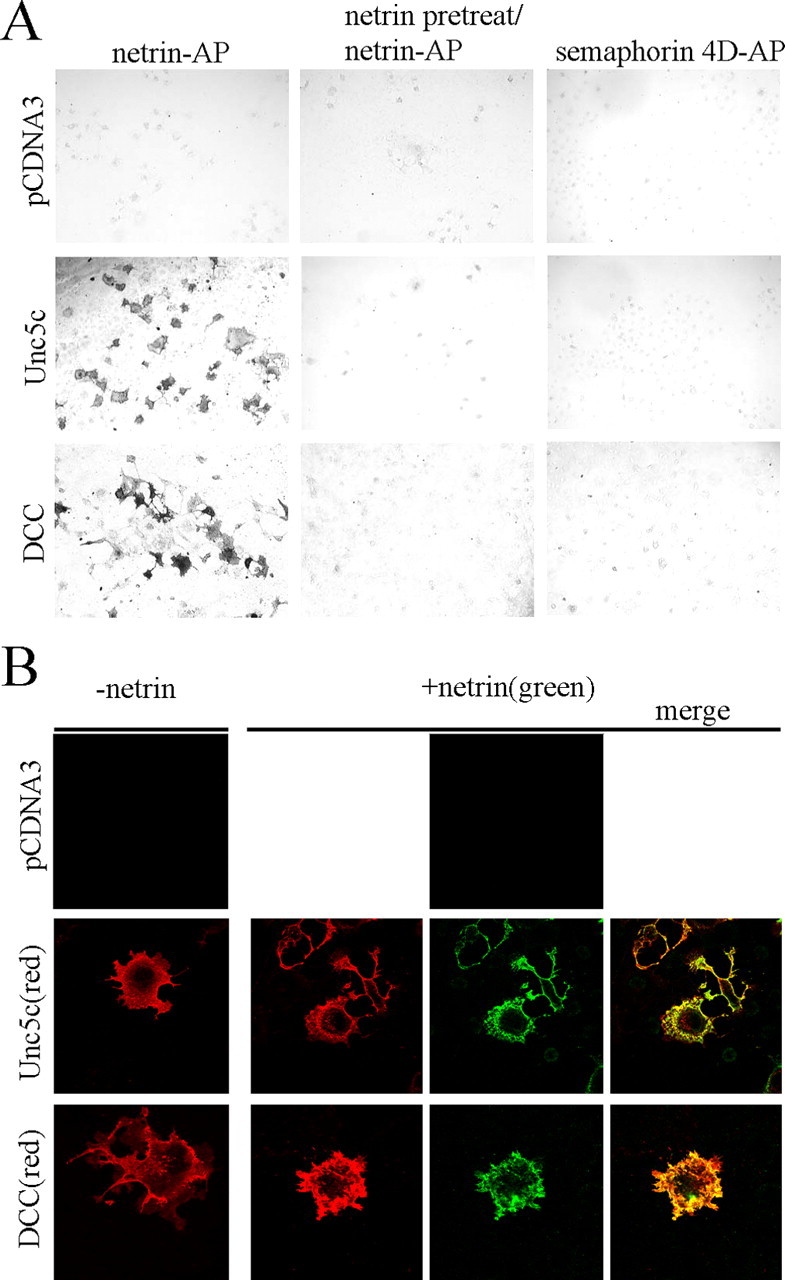
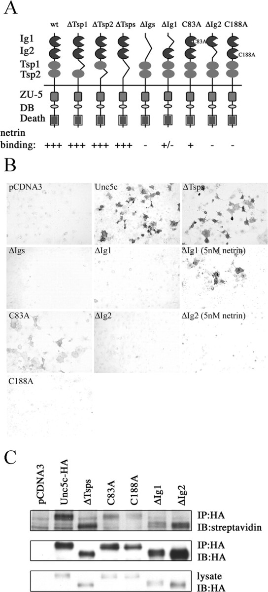
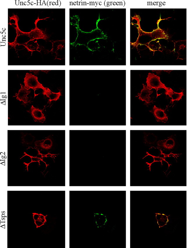
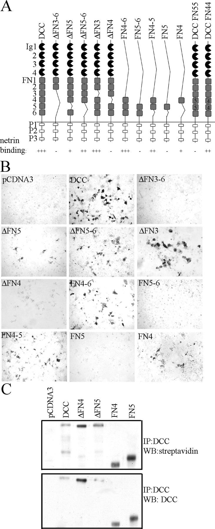
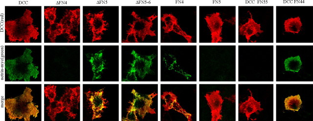
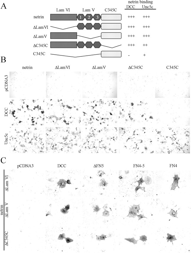
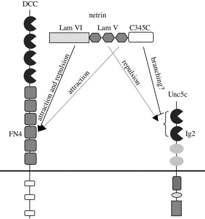
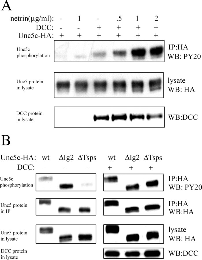
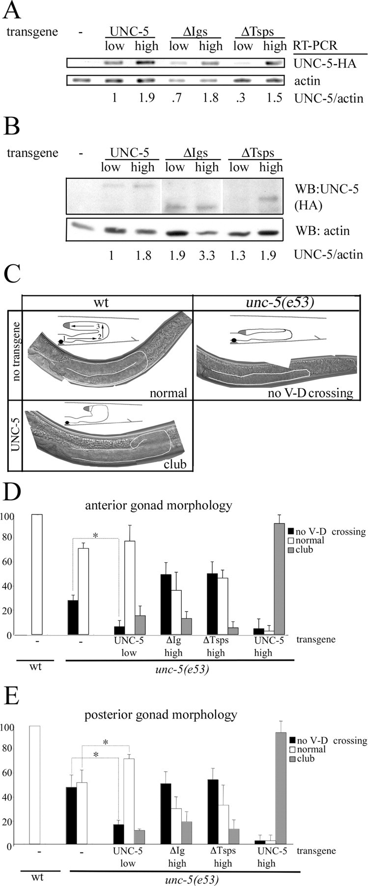
Netrin and its receptors Unc5 and deleted in colorectal carcinoma (DCC) regulate axon guidance and cell migration. We defined domains involved in the interactions between netrin-1, DCC, and Unc5c. We show that Unc5 requires both Ig domains to interact with netrin. DCC binds through the fourth fibronectin type III domain, whereas netrin binds through multiple domains to both receptors. We examined the functional consequences of removing the netrin binding and nonbinding domains from Unc5 in vitro and in vivo. In human embryonic kidney 293 cells, removal of the netrin binding second Ig domain causes an increase in basal tyrosine phosphorylation, whereas removal of the netrin nonbinding thrombospondin domains decreases tyrosine phosphorylation. Moreover, experiments in Caenorhabditis elegans indicate that both netrin binding and nonbinding domains are necessary for phenotypic rescue of an unc-5 loss of function mutation.


Figures

Figure 1.
Figure 1.
A, Binding of netrin-AP to Unc5c- and DCC-expressing Cos-7 cells, but not pCDNA3 transfected cells (left column). Preincubation of cells with purified chicken netrin blocks netrin-AP binding (middle column). No binding is observed between semaphorin 4D-AP and DCC- and Unc5c-expressing cells (right column). B, Colocalization of myc-netrin bound to DCC- and Unc5-expressing Cos-7 cells by immunofluorescence confocal microscopy. Examples of netrin-bound cells are shown for Unc5 (second row) and DCC (third row) with and without incubation of netrin before fixation. Unc5c and DCC (red) show surface expression. Bound netrin (green) is seen at the surface of DCC- or Unc5c-expressing cells only. No netrin binding is observed to untransfected cells. The merged image (last column) shows colocalization (yellow) of netrin bound to its receptors.
Figure 2.
Figure 2.
A, Unc5 has two Ig domains, two Tsp domains, a ZO-1/Unc5 (ZU-5) domain, a DCC binding motif (DB), and a death domain. Binding is summarized as follows to Unc5c and deletion constructs: (+++) strong, (+) weak, (+/-) detectable above background at 5 nm, and (-) none detected. B, Binding of Unc5c and deletion mutant expressing Cos-7 cells to netrin-AP. Dark cells are Unc5c-expressing cells that have bound netrin-AP. C, Cell surface expression of Unc5c ectodomain deletions and point mutants. The top panel shows biotinylated proteins in the IP, the second shows protein in the IP, and the third protein in the lysates. All proteins show incorporation of the cell-impermeable biotinylation reagent.
Figure 3.
Figure 3.
Binding and colocalization of Unc5c and deletion mutants to myc-tagged netrin. Red indicates Unc5 expression. Green shows netrin binding. The merged image shows colocalization of netrin to Unc5c and Unc5c deletion constructs. The results show a pattern similar to netrin-AP staining (Fig. 2 B).
Figure 4.
Figure 4.
A, DCC has four Ig domains, six fibronectin type III (FN), and three conserved motifs P1, P2, and P3. Binding to DCC and deletion mutants is summarized as follows: (+++) strong, (++) moderate, (+) weak, and (-) none detected. A, B, Binding of DCC-expressing Cos-7 cells to netrin-AP. C, Evidence of surface expression of DCC deletion constructs. Cell surface expression is indicated by the incorporation of a cell-impermeable biotinylation reagent as detected by streptavidin-HRP in Western analysis. All constructs show evidence of surface expression.
Figure 5.
Figure 5.
Binding and colocalization of DCC and deletion mutants with myc-tagged netrin. Red indicates DCC expression. Green shows bound netrin. The merged image shows colocalization of netrin with DCC and deletion constructs. The results show the same pattern of binding as with netrin-AP staining (Fig. 4 B).
Figure 6.
Figure 6.
A, Netrin has three domains, a Laminin VI-like domain, three EGF repeats constituting a Laminin V-like domain, and a C-terminal C345C domain. Qualitative assessment of binding with netrin and deletion constructs is as follows: (+++) strong, (++) moderate, (+) weak, and (-) none detected. B, Binding of DCC- and Unc5c-expressing Cos-7 cells to netrin-AP deletion mutants. C, Netrin-AP deletion constructs binding to DCC deletions shows that multiple netrin domains interact with DCC through FN4. Because these are weak interactions, individual netrin-bound cells are shown. All netrin deletion constructs show binding to ΔFN5 and FN4. FN4-5 does not differ from the binding seen to FN4.
Figure 7.
Figure 7.
A model of Unc5, DCC, and netrin interactions. Dotted lines denote inferences from the work of Lim and Wadsworth (2002) regarding particular netrin EGF-like repeats involved in receptor binding. Critical domains in DCC (FN4) and Unc5 (Ig2) are indicated. A bracket is used to note Ig1 of Unc5, which also contributes to netrin binding, but is insufficient by itself to bind netrin in our assays.
Figure 8.
Figure 8.
Basal and netrin-stimulated tyrosine phosphorylation of Unc5c in HEK293 cells. A, Phosphorylation of full-length Unc5c. Unc5c phosphorylation is stimulated by netrin and augmented by DCC coexpression. DCC coexpression and netrin stimulation have additive effects. B, Phosphorylation of Unc5c and deletion constructs. ΔIg2 shows more basal tyrosine phosphorylation, and ΔTsps shows less tyrosine phosphorylation than full-length Unc5c. The Unc5c deletion constructs show similar phosphorylation when DCC is coexpressed.
Figure 9.
Figure 9.
A, RT-PCR from RNA purified from nematodes expressing UNC-5 transgenes. The intensity of the transgenic UNC-5-specific band compared with the actin control indicates the relative amounts of transcription of each transgene. A reverse primer embedded in the HA tag was used to ensure transgene specificity. B, Fifteen transgenic worms for each strain were collected and analyzed for UNC-5-HA expression by Western blot analysis. The ratio of UNC-5 to actin is calculated. C, Images showing the posterior gonad arm in wt and unc-5(e53) worms. In wt worms, U-shaped gonad arms result from the migration of DTCs. The three phases of this migration in wt and unc-5 worms are depicted in the diagram inserts with numbered arrows. The second phase of this migration, in which DTCs move from the ventral side to the dorsal side, is dependent on UNC-6/netrin, UNC-40/DCC, and UNC-5. Representative phenotypes are shown for the posterior gonad arm (outlined in white) for each wt, unc-5(e53), and a worm expressing the UNC-5 transgene at the higher expression level. In the unc-5(e53) panel, the DTC does not have a ventral to dorsal migration and returns to the vulval region in a different plane of focus than the first phase of migration. In all pictures, ventral is at bottom and posterior to the right. D, E, Morphologies of the anterior (D) and posterior (E) gonad arms with UNC-5 transgene expression. In the development of unc-5(e53) worms, DTCs frequently fail to have a ventral to dorsal migration (black bars). With transgenic expression of UNC-5 at the lower level this percentage decreases, indicating phenotypic rescue (*p < 0.01). UNC-5ΔIgs, UNC-5ΔTsps, or UNC-5 expressed at the higher level did not rescue the DTC migration defect. The club-shaped gain-of-function phenotype is also observed at different frequencies between constructs (gray bars). White bars indicate normal gonad formation.

References

    1. Bennett KL, Bradshaw J, Youngman T, Rodgers J, Greenfield B, Aruffo A, Linsley PS (1997) Deleted in colorectal carcinoma (DCC) binds heparin via its fifth fibronectin type III domain. J Biol Chem 272: 26940-26946. - PubMed
    1. Chan SS, Zheng H, Su MW, Wilk R, Killeen MT, Hedgecock EM, Culotti JG (1996) UNC-40, a C. elegans homolog of DCC (Deleted in Colorectal Cancer), is required in motile cells responding to UNC-6 netrin cues. Cell 87: 187-195. - PubMed
    1. Colamarino SA, Tessier-Lavigne M (1995) The axonal chemoattractant netrin-1 is also a chemorepellent for trochlear motor axons. Cell 81: 621-629. - PubMed
    1. Colavita A, Krishna S, Zheng H, Padgett RW, Culotti JG (1998) Pioneer axon guidance by UNC-129, a C. elegans TGF-beta. Science 281: 706-709. - PubMed
    1. Dickson B (2002) Molecular mechanisms of axon guidance. Science 298: 1959-1964. - PubMed

Publication types

MeSH terms

LinkOut - more resources