Comparative Study
. 2005 Feb;16(2):776-93.
doi: 10.1091/mbc.e04-08-0700. Epub 2004 Dec 1.

Synthetic genetic array analysis of the PtdIns 4-kinase Pik1p identifies components in a Golgi-specific Ypt31/rab-GTPase signaling pathway

Affiliations
Comparative Study

Synthetic genetic array analysis of the PtdIns 4-kinase Pik1p identifies components in a Golgi-specific Ypt31/rab-GTPase signaling pathway

Vicki A Sciorra et al. Mol Biol Cell. 2005 Feb.

Abstract

Phosphorylated derivatives of phosphatidylinositol are essential regulators of both endocytic and exocytic trafficking in eukaryotic cells. In Saccharomyces cerevisiae, the phosphatidylinositol 4-kinase, Pik1p generates a distinct pool of PtdIns(4)P that is required for normal Golgi structure and secretory function. Here, we utilize a synthetic genetic array analysis of a conditional pik1 mutant to identify candidate components of the Pik1p/PtdIns(4)P signaling pathway at the Golgi. Our data suggest a mechanistic involvement for Pik1p with a specific subset of Golgi-associated proteins, including the Ypt31p rab-GTPase and the TRAPPII protein complex, to regulate protein trafficking through the secretory pathway. We further demonstrate that TRAPPII specifically functions in a Ypt31p-dependent pathway and identify Gyp2p as the first biologically relevant GTPase activating protein for Ypt31p. We propose that multiple stage-specific signals, which may include Pik1p/PtdIns(4)P, TRAPPII and Gyp2p, impinge upon Ypt31 signaling to regulate Golgi secretory function.


Figures
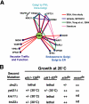
Figure 1.
Figure 1.
Phosphoinositide kinase specific genetic interactions. (A) Schematic of genes identified by SGA involved in membrane trafficking with known genetic interactions (synthetic sick or lethal). (B) Genetic interactions of four trafficking genes obtained from SGA (drs2Δ, ypt31Δ, kre11Δ, and trs33Δ) were analyzed in pik1-139ts, pik1–83ts, stt4ts, and mss4ts. Tetrads were isolated at 26°C and the growth of the double mutants was assessed at 26°C. +/-; reduced growth, ++; similar to growth of single mutants. Temperature at which lethality is observed is given in parentheses.
Figure 2.
Figure 2.
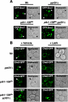
Trafficking of Snc1p and Chs3p requires Pik1p and Ypt31p. (A) GFP-Snc1p or (B) Chs3p-GFP distribution at 26°C in live cells, either wild-type (Wt) or carrying the indicated mutation, ypt31Δ, pik1-139ts, and pik1-139ts ypt31Δ. Arrows indicate bud necks and arrowheads indicate bud tips. (B) Chs3p-GFP transport to the cell surface was monitored by blocking endocytosis with LatA (200 μM, Molecular Probes) or treated with vehicle (dimethyl sulfoxide) as described in Valdivia et al. (2002).
Figure 3.
Figure 3.
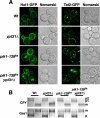
Trafficking competence of pik1-139ts ypt31Δ double mutant cells at permissive temperature. (A) Hxt1p-GFP and Tat2p-GFP distribution at 26°C in Wt, ypt31Δ, pik1-139ts, and pik1-139ts ypt31Δ. (B) Indicated yeast strains (Wt, ypt31Δ, pik1-139ts, and pik1-139ts ypt31Δ) were metabolically labeled with Tran-35S for 10 min, and chased in the presence of excess methionine/cysteine for 30 min at 26°C. CPY and Gas1p were immunoprecipitated from cell lysates, resolved by SDS-PAGE, and visualized by fluorography. p, precursor form; m, mature form.
Figure 4.
Figure 4.
Pik1p and Ypt31p act in similar biological pathways. (A) Temperature-sensitive growth of yeast expressing ypt31-101ts in ypt32Δ background is rescued by expressing low-copy YPT31. (B and C) Wt, ypt32Δ, and ypt32Δypt31-101ts double mutant cells were incubated at the indicated temperature for 10 or 30 min, metabolically labeled with Tran-35S for 10 min, and chased in the presence of excess methionine/cysteine for 30 min. CPY and Gas1p were analyzed as described in the legend to Figure 3. To assay for general secretion competence, cells and media were separated by centrifugation, and proteins secreted into the media during the pulse-chase were visualized by SDS-PAGE and fluorography. (D) ypt32Δypt31-101ts double mutant cells expressing GFP-Snc1p were grown to early/midlog phase, shifted to 26°C or 38°C for 30 min, and observed by fluorescence microscopy.
Figure 5.
Figure 5.
Phosphoinositide levels in Wt, ypt32Δ, and ypt32Δypt31-101ts double mutant cells. Cells were incubated at the temperature indicated for 10 min and labeled with myo-[2-3H]inositol for 45 min, and phosphoinositides were analyzed as previously described in Rudge et al. (2004). The levels of each indicated phosphoinositide are expressed as a percentage of the total 3H-labeled phosphoinositides analyzed by HPLC and represent the average of two independent experiments done in duplicate (n = 4).
Figure 6.
Figure 6.
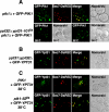
Independent localization of Pik1p and Ypt31p to the trans-Golgi network. (A) pik1Δ cells expressing GFP-Pik1p and Sec7p-DsRed were examined by fluorescence microscopy at 26°C. ypt32Δypt31-101tspik1Δ cells expressing GFP-Pik1p were grown at 26°C or shifted for 1 h at 38°C and examined by fluorescence microscopy. (B) ypt31Δ ypt32Δ cells expressing GFP-Ypt31p and Sec7p-DsRed were grown at 26°C and examined by fluorescence microscopy. (C) Wild-type (PIK1), pik1–83ts, or pik1-139ts (unpublished data) cells expressing GFP-YPT31 from a low copy plasmid and Sec7p-DsRed were grown at 26°C (unpublished data) or shifted 1 h at 38°C and examined by fluorescence microscopy.
Figure 7.
Figure 7.
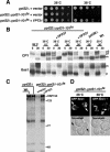
GTP-dependent suppression of TRAPPII lethality and trafficking defects by YPT31. (A) Tetrad analysis of diploid strains were transformed with high-copy wild-type YPT31, or ypt31 Q72L, sporulated, and dissected on YPD plates at 26°C. Haploid cells with deleted gene(s) carrying either YPT31 or ypt31 Q72L are circled (confirmed by PCR and growth on selection media). (B) trs130-HAts (33 aa deleted from C-terminus of TRS130, and tagged with HA) was examined for temperature-sensitive growth when expressing low (cen) or high (2 μ) copies of YPT31 or ypt31 Q72L. (C) Wt, TRS130-HA, or trs130-HAts cells expressing vector, YPT31 or ypt31 Q72L, were incubated at 26 or 38°C for 30 min, metabolically labeled with Tran-35S for 10 min, and chased in the presence of excess methionine/cysteine for 30 min. CPY and GAS1p were analyzed as described in the legend to Figure 3. (D) General secretion competence was analyzed as described in the legend to Figure 4.
Figure 8.
Figure 8.
Gyp2p is a GAP for Ypt31p in vivo. (A) gyp2 deletion suppresses the temperature-sensitive growth of trs130-HAts. (B) SEC4 and YPT6 (high-copy) do not suppress the temperature-sensitive growth defect of trs130-HAts. (C) gyp2 deletion suppresses the temperature-sensitive growth of ypt32Δypt31-101ts double mutant cells. (D) gyp2 deletion specifically rescues trafficking defects of ypt32Δypt31-101ts double mutant cells. ypt32Δypt31-101ts, ypt32Δypt31-101ts gyp1Δ, and ypt32Δypt31-101tsgyp2Δ were incubated at 37°C for 30 min, metabolically labeled with Tran-35S for 10 min, and chased in the presence of excess methionine/cysteine for the indicated minutes. CPY and Gas1p were analyzed as described in the legend to Figure 3.
Figure 9.
Figure 9.
Gyp2p requires its GRAM domain and GAP activity for function in cells. (A) Schematic representation of the conserved domains in Gyp2 family in human, Drosophila, C. elegans, and yeast. EfH, EfHand; cc, coiled-coil. (B) Serial dilution of yeast cells expressing wild-type or mutants of GYP2 from low-copy plasmids at 26 and 37°C. (C) trs130-HAts gyp2Δ cells expressing GFP-GYP2, GFP-gyp2 G80E, or GFP-gyp2 R295K from low-copy plasmids were grown at 26°C and shifted to 37°C for 1 h, and total cellular protein content were TCA-precipitated and resolved by Western blotting. Glucose 6-phosphate dehydrogenase (G6PDH) was examined as a loading control.
Figure 10.
Figure 10.
(A) Snc1p trafficking to the plasma membrane (PM) in yeast requires Golgi and early endosome (EE) function. Inactivation of Pik1p or Ypt31p causes Snc1p to accumulate intracellularly. We propose that the primary role for Pik1p/PtdIns(4)P is in maintaining the structural integrity and molecular identity of the trans-Golgi and that unidentified multivalent adaptor proteins may be recruited by Pik1p/PtdIns(4)P and/or activated Ypt31/32p for the coordination of cargo-loading, budding, and/or fission at the trans-Golgi. Alternatively, Ypt31/32p may also participate in retrograde traffic of recycled cargoes by regulating early endosome-to-Golgi transport. (B) The orderly cascade of yeast exocytic rab-GTPases. The cis-Golgi rab-GTPase, Ypt1p and the multicomponent complex, TRAPPII act upstream to the trans-Golgi rab-GTPase, Ypt31p, possibly leading to its activation. Activated, GTP-bound Ypt31p interacts with Sec2p, the GEF for Sec4p (Ortiz et al., 2002), whereas activated-Ypt6p may recruit Gyp2p (Siniossoglou and Pelham, 2001), which inactivates Ypt31p, allowing the rab-GTPase cycle of activation/inactivation at the Golgi to continue.

Similar articles

Cited by

References

    1. Albert, S., and Gallwitz, D. (1999). Two new members of a family of Ypt/Rab GTPase activating proteins. J. Biol. Chem. 274, 33186-33189. - PubMed
    1. Albert, S., Will, E., and Gallwitz, D. (1999). Identification of the catalytic domains and their functionally critical arginine residues of two yeast GTPase-activating proteins specific for Ypt/Rab transport GTPases. EMBO J. 18, 5216-5225. - PMC - PubMed
    1. Audhya, A., Foti, M., and Emr, S. D. (2000). Distinct roles for the yeast phosphoinositol 4-kinases, Stt4p and Pik1p, in secretion, cell growth, and organelle membrane dynamics. Mol. Biol. Cell 11, 2673-2689. - PMC - PubMed
    1. Audhya, A., and Emr, S. D. (2002). Stt4 PI4-kinase localizes to the plasma membrane and functions I the Pkc1-mediated MAP kinase cascade. Dev. Cell 2, 593-605. - PubMed
    1. Begley, M. J., Taylor, G. S., Kim, S.-A., Veine, D. M., Dixon, J. E., and Stuckey, J. A. (2003). Crystal structure of a phosphoinsitide phosphatase, MTMR 2, insights into myotubular myopathy and Charcot-Marie Tooth Syndrome. Mol. Cell 12, 1391-1402. - PubMed

Publication types

MeSH terms