Comparative Study
. 2004 Dec;70(12):7110-8.
doi: 10.1128/AEM.70.12.7110-7118.2004.

Streptomyces coelicolor A3(2) lacks a genomic island present in the chromosome of Streptomyces lividans 66

Affiliations
Comparative Study

Streptomyces coelicolor A3(2) lacks a genomic island present in the chromosome of Streptomyces lividans 66

Xiufen Zhou et al. Appl Environ Microbiol. 2004 Dec.

Abstract

Streptomyces lividans ZX1 has become a preferred host for DNA cloning in Streptomyces species over its progenitor, the wild-type strain 66 (stock number 1326 from the John Innes Center collection), especially when stable DNA is crucial for in vitro electrophoresis, because DNA from strain 66 contains a novel modification that makes it sensitive to oxidative double-strand cleavage during electrophoresis. Detailed analysis of this modification-deficient mutant (ZX1) revealed that it has several additional phenotypic traits associated with a chromosomal deletion of ca. 90 kb, which was cloned and mapped by using a cosmid library. Comparative sequence analysis of two clones containing the left and right deletion ends originating from strain 66 and one clone with the deletion and fused sequence cloned from strain ZX1 revealed a perfect 15-bp direct repeat, which may have mediated deletion and fusion to yield strain ZX1 by site-specific recombination. Analysis of AseI linking clones in the deleted region in relation to the published AseI map of strain ZX1 yielded a complete AseI map for the S. lividans 66 genome, on which the relative positions of a cloned phage phiHAU3 resistance (phiHAU3r) gene and the dnd gene cluster were precisely localized. Comparison of S. lividans ZX1 and its progenitor 66, as well as the sequenced genome of its close relative, Streptomyces coelicolor M145, reveals that the ca. 90-kb deletion in strain ZX1 may have originated from an insertion from an unknown source.


Figures

FIG. 1.
FIG. 1.
Summary of the phenotypic differences between S. lividans JT46 and ZX1. (A) Dnd+ phenotype (sensitizing DNA to degradation during electrophoresis) in strain JT46 and Dnd phenotype in strain ZX1. Lane M contains S. coelicolor M145 digested with AseI used as size markers. (B) φHAU3 resistance exhibited by JT46 versus φHAU3 sensitivity exhibited by ZX1. (C) Good sporulation in JT46 but poor sporulation in ZX1. (D) Good melC expression in JT46 but poor melC expression in ZX1. (E) Transformation frequency of ZX1 is ca. 10 times higher than that of JT46.
FIG. 2.
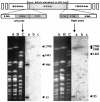
FIG. 2.
(Left) Comparison of the AseI banding patterns of S. lividans 66 and ZX1 by PFGE. Strain 66 is shown as 1326 in the figure. The white arrows point to bands seen in strain 66 but not in strain ZX1. The solid black arrows point to a band seen in ZX1 after deletion and fusion but not seen in strain 66 and a 52-kb band corresponding to the linear plasmid SLP2 present in strain 66. AseI-digested M145 DNA was run in parallel as size markers; the sizes (in kilobases) derived from genome sequences (3) are indicated at the sides of the gel. (Right) Determination of the terminal AseI fragments of S. lividans 66 bound with protein. Samples treated with proteinase K (+P) or not treated with proteinase K (−P) were digested with AseI and separated on a 1% agarose gel with a 90-s pulse for 20 h, followed by a 220-s pulse for 25 h, at 4.5 V/cm, in 0.5× TBE buffer. White arrows point to the AseI fragments affected by proteinase K treatment, and certain AseI fragments unaffected by proteinase K treatment are indicated by solid black arrows. Lane M contains S. coelicolor M145 digested with AseI used as size markers (in kilobases).
FIG. 3.
FIG. 3.
Physical maps of S. lividans 66 and ZX1. Strain 66 is shown as 1326 in the figure. The region present in strain 66 but not present in strain ZX1 is enlarged to show overlapping cosmids ordered by T3 and T7 riboprobes. The positions of the dnd gene cluster (responsible for the Dnd phenotype of S. lividans) and the φHAU3r gene (conferring phage φHAU3 resistance on S. lividans) (37) are shown as black boxes. The positions of two genes immediately flanking the left deletion junction, ORF1 (a putative phage integrase) and ORF2 (a putative transposase) are indicated by the small black triangles facing left. The ORF1 and ORF2 sequences have been assigned accession number AY626162. Vertical broken lines represent BamHI sites and deletion junctions in DNA. c. 90 kb, ca. 90 kb.
FIG. 4.
FIG. 4.
Precise localization of the deleted region of ca. 90 kb (shaded) on the physical map of S. lividans 66. (Top) Schematic representation of the relationship of the regions flanking the ca. 90-kb deletion, with enlarged regions used as probes for the localization of the left junction (left probe) from the linking cosmid 16H2 and right junction (right probe) from the linking cosmid 16C3. (Bottom) Results of Southern blots of PFGE of samples of S. lividans and S. coelicolor. AseI-digested samples of S. lividans 66 (lane A), S. lividans ZX1 (lane B), and S. coelicolor M145 (lane C) (used as size markers [in kilobases]), hybridized using the left or right probe to give autoradiographic signals (lanes a, b, and c) corresponding to lanes A, B, and C. Samples were separated on a 1% agarose gel with a 90-s pulse for 20 h, followed by a 220-s pulse for 25 h, at 4.5 V/cm, in 0.5× TBE buffer.
FIG. 5.
FIG. 5.
Sequencing analysis of the deleted and fused sequences and implication of a foreign insertion in S. lividans 66. Strain 66 is shown as 1326 in the figure. (Top) Sequences of plasmids pHZ1908, pHZ1906b, and pHZ1906a. Sequencing of a 1.4-kb SalI fragment in pHZ1908 (carrying the left insertion junction), a 3-kb BglII fragment in pHZ1906a (carrying the right insertion junction) cloned from strain 66 genome, and a 0.8-kb SstII-SalI fragment in pHZ1906b (carrying the deletion and the fused sequence) cloned from the S. lividans ZX1 genome revealed 15-bp direct repeat sequences. The 15-bp direct repeat sequences are shown shaded. The 15-bp direct repeat sequences probably mediate the deletion and fusion that form strain ZX1 by site-specific recombination. (Middle) Hypothetical evolution of S. lividans 66 from a strain with a genome corresponding to that of ZX1 by the acquisition of the deleted region (ca. 90 kb) from an unknown source(s). The corresponding portions of the DNA sequences before (66 [1326]) and after (ZX1) deletion and the relevant part of the S. coelicolor M145 DNA sequence, which is identical to the ZX1 region after deletion and fusion, are aligned. The perfect 15-bp direct repeat, which probably mediates the insertion of a foreign sequence by site-specific recombination, is indicated by white letters in black boxes (the first three of the highlighted nucleotides conform to the nucleotides in the plus strand of the stop codon for SCO5998 [murA1], transcribing from right to left). The relative positions of genes in S. coelicolor M145 flanking the putative insertion, SCO5997 on the left and SCO5998 (murA1) and SCO5999 on the right, are shown inside the AseI-B fragment. (Bottom) Relative positions of the 15-bp direct repeats detected (small arrows) and comparison of the architecture of the genes flanking the direct repeats in S. avermitilis. The SAV2661 and SAV2662 ORFs are located between the two 15-bp direct repeats. The SAV2663 and SAV2259 ORFS flank both sides of the 15-bp direct repeat sequence. These ORFs were not related to any other genes flanking the 15-bp sequence apart from murA1. The comparison of the architecture of the genes showed that strain M145 was related to strains ZX1 and 66; the homologous murA1 gene contained a nucleotide sequence identical to the sequence of one of the 15-bp direct repeats in the same position.

References

    1. Alting-Mees, M. A., and J. M. Short. 1989. pBluescript II: gene mapping vectors. Nucleic Acids Res. 17:9494. - PMC - PubMed
    1. Bao, K., Z. Hu, X. Zhou, Q. Zhou, and Z. Deng. 1997. A bifunctional cosmid vector for the mobilized conjugal transfer of DNA from E. coli to Streptomyces sp. Prog. Nat. Sci. 7:568-572.
    1. Bentley, S. D., K. F. Chater, A. M. Cerdeno-Tarraga, G. L. Challis, N. R. Thomson, K. D. James, D. E. Harris, M. A. Quail, H. Kieser, D. Harper, A. Bateman, S. Brown, G. Chandra, C. W. Chen, M. Collins, A. Cronin, A. Fraser, A. Goble, J. Hidalgo, T. Hornsby, S. Howarth, C. H. Huang, T. Kieser, L. Larke, L. Murphy, K. Oliver, S. O'Neil, E. Rabbinowitsch, M. A. Rajandream, K. Rutherford, S. Rutter, K. Seeger, D. Saunders, S. Sharp, R. Squares, S. Squares, K. Taylor, T. Warren, A. Wietzorrek, J. Woodward, B. G. Barrell, J. Parkhill, and D. A. Hopwood. 2002. Complete genome sequence of the model actinomycete Streptomyces coelicolor A3(2). Nature 417:141-147. - PubMed
    1. Bierman, M., R. Logan, K. O'Brien, E. T. Seno, R. N. Rao, and B. E. Schoner. 1992. Plasmid cloning vectors for the conjugal transfer of DNA from Escherichia coli to Streptomyces spp. Gene 116:43-49. - PubMed
    1. Borck, K., J. D. Beggs, W. J. Brammar, A. S. Hopkins, and N. E. Murray. 1976. The construction in vitro of transducing derivatives of phage lambda. Mol. Gen. Genet. 146:199-207. - PubMed

Publication types

MeSH terms

Substances

LinkOut - more resources