. 2004 Dec;70(12):7418-25.
doi: 10.1128/AEM.70.12.7418-7425.2004.

Hypothesis for the role of nutrient starvation in biofilm detachment

Affiliations

Hypothesis for the role of nutrient starvation in biofilm detachment

Stephen M Hunt et al. Appl Environ Microbiol. 2004 Dec.

Abstract

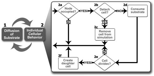
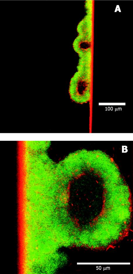
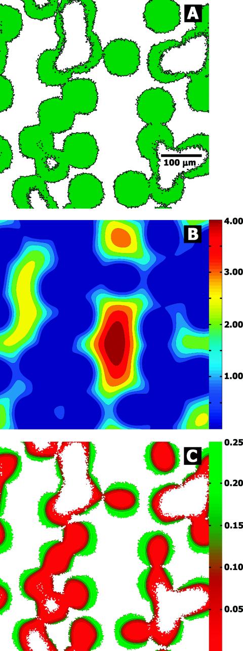
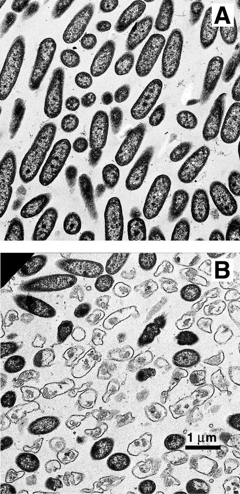
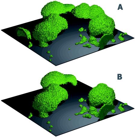
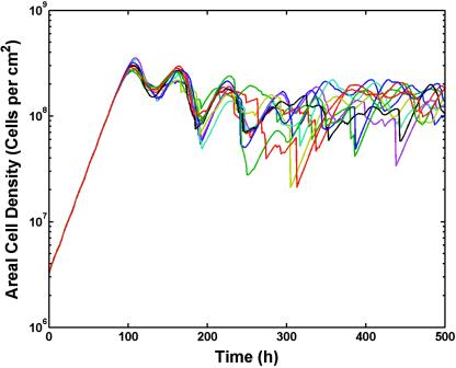
A combination of experimental and theoretical approaches was used to investigate the role of nutrient starvation as a potential trigger for biofilm detachment. Experimental observations of detachment in a variety of biofilm systems were made with pure cultures of Pseudomonas aeruginosa. These observations indicated that biofilms grown under continuous-flow conditions detached after flow was stopped, that hollow cell clusters were sometimes observed in biofilms grown in flow cells, and that lysed cells were apparent in the internal strata of colony biofilms. When biofilms were nutrient starved under continuous-flow conditions, detachment still occurred, suggesting that starvation and not the accumulation of a metabolic product was responsible for triggering detachment in this particular system. A cellular automata computer model of biofilm dynamics was used to explore the starvation-dependent detachment mechanism. The model predicted biofilm structures and dynamics that were qualitatively similar to those observed experimentally. The predicted features included centrally located voids appearing in sufficiently large cell clusters, gradients in growth rate within these clusters, and the release of most of the biofilm with simulated stopped-flow conditions. The model was also able to predict biofilm sloughing resulting solely from this detachment mechanism. These results support the conjecture that nutrient starvation is an environmental cue for the release of microbes from a biofilm.


Figures

FIG. 1.
FIG. 1.
General procedure followed in a typical BacLAB simulation. See the text for an explanation of the sequence of operations.
FIG. 2.
FIG. 2.
Biofilm detachment after stopping of the flow of medium in a drip-flow reactor (circles) and as predicted by BacLAB (average of six simulations) (solid line). Approximately 90% of the biomass detached after 1 day under static conditions.
FIG. 3.
FIG. 3.
Confocal laser scanning microscopy of hollow P. aeruginosa biofilm cell clusters. Biofilms were grown in glass capillary tubes under continuous flow. The bacteria contained green fluorescent protein and appeared green. The specimen was counterstained with rhodamine B (red), the primary utility of which was in locating the glass wall. Shown are two different locations of the same specimen.
FIG. 4.
FIG. 4.
Representative BacLAB simulation predicting hollow biofilm cell clusters. All three panels show patterns at the base of the biofilm across the square substratum area. The distribution of biomass at the substratum (green in panel A) suggests that larger clusters develop hollow interiors. Also shown are the predicted distributions of oxygen concentrations, in milligrams liter−1, at the substratum (B) and the specific growth rates, in hours−1, at the substratum (C). These simulations predicted sharp gradients in the concentration of the metabolic substrate (B) and the growth status of cells in the biofilm (C).
FIG. 5.
FIG. 5.
Transmission electron microscopic analysis of biofilms. (A) Little lysis is evident in cells near the air interface of the biofilm. (B) Cell lysis is observed in the interior of P. aeruginosa colony biofilms.
FIG. 6.
FIG. 6.
BacLAB simulation showing biofilm sloughing. Biofilm structures at 235 h (A), before the sloughing event, and at 240 h (B), after the sloughing event, are shown. An entire cell cluster near the rear corner of the simulated area disappeared in the interval between the two time points. (The entire simulation can be viewed at http://www.erc.montana.edu/Res-Lib99-SW/Movies/Database/MD_DisplayScript.asp.)
FIG. 7.
FIG. 7.
BacLAB replicate simulations showing sloughing as revealed by sharp decreases in areal cell density. The parameter settings were identical in these 10 simulations; only the random initial distributions of cells on the substrata differed in the 10 runs. Sloughing events, defined as a loss of 50% of biofilm biomass in a single time step (1 h), occurred in 6 of the 10 simulations.

References

    1. Allison, D. G., B. Ruiz, C. SanJose, A. Jaspe, and P. Gilbert. 1998. Extracellular products as mediators of the formation and detachment of Pseudomonas fluorescens biofilms. FEMS Microbiol. Lett. 167:179-184. - PubMed
    1. Applegate, D. H., and J. D. Bryers. 1991. Effects of carbon and oxygen limitations and calcium concentrations on biofilm removal processes. Biotechnol. Bioeng. 37:17-25. - PubMed
    1. Boyd, A., and A. M. Chakrabarty. 1994. Role of alginate lyase in cell detachment of Pseudomonas aeruginosa. Appl. Environ. Microbiol. 60:2355-2359. - PMC - PubMed
    1. Caccavo, F., B. Frolund, F. V. Kloeke, and P. H. Nielsen. 1996. Deflocculation of activated sludge by the dissimilatory Fe(III)-reducing bacterium Shewanella alga BrY. Appl. Environ. Microbiol. 62:1487-1490. - PMC - PubMed
    1. Chang, H. T., B. E. Rittmann, D. Amar, R. Heim, O. Ehlinger, and Y. Lesty. 1991. Biofilm detachment mechanisms in a liquid-fluidized bed. Biotechnol. Bioeng. 38:499-506. - PubMed

LinkOut - more resources