Review
. 2004 Nov;205(5):335-47.
doi: 10.1111/j.0021-8782.2004.00345.x.

Evolution of the vertebrate jaw: comparative embryology and molecular developmental biology reveal the factors behind evolutionary novelty

Affiliations
Review

Evolution of the vertebrate jaw: comparative embryology and molecular developmental biology reveal the factors behind evolutionary novelty

Shigeru Kuratani. J Anat. 2004 Nov.

Abstract

It is generally believed that the jaw arose through the simple transformation of an ancestral rostral gill arch. The gnathostome jaw differentiates from Hox-free crest cells in the mandibular arch, and this is also apparent in the lamprey. The basic Hox code, including the Hox-free default state in the mandibular arch, may have been present in the common ancestor, and jaw patterning appears to have been secondarily constructed in the gnathostomes. The distribution of the cephalic neural crest cells is similar in the early pharyngula of gnathostomes and lampreys, but different cell subsets form the oral apparatus in each group through epithelial-mesenchymal interactions: and this heterotopy is likely to have been an important evolutionary change that permitted jaw differentiation. This theory implies that the premandibular crest cells differentiate into the upper lip, or the dorsal subdivision of the oral apparatus in the lamprey, whereas the equivalent cell population forms the trabecula of the skull base in gnathostomes. Because the gnathostome oral apparatus is derived exclusively from the mandibular arch, the concepts 'oral' and 'mandibular' must be dissociated. The 'lamprey trabecula' develops from mandibular mesoderm, and is not homologous with the gnathostome trabecula, which develops from premandibular crest cells. Thus the jaw evolved as an evolutionary novelty through tissue rearrangements and topographical changes in tissue interactions.


Figures
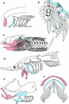
Fig. 1
Fig. 1
Visceral skeletal systems in various gnathostomes. Visceral skeletons of Chimaera monstrosa (A: Holocephali), Callorhynchus (B: Holocephali), Chlamidoselachus (C: Elasmobranchii), Acipenser sturio (D: Chondrostei), Gasterosteus aculeatus (E: Teleostei) and Triron cristatus (F: Urodela) are shown. Mandibular arches are coloured pink, the hyoid arch light blue, and the more posterior respiratory arches (real branchial arches) grey. Note that, in each gnathostome species shown here, mandibular and hyoid arches are morphologically differentiated to take distinct shapes, whereas the branchial arches look similar to each other. Redrawn from Edgeworth (1935) (A, B, E, F) and Gregory (1933) (C and D).
Fig. 2
Fig. 2
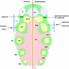
Pharyngeal anatomy of the ammocoete larva of the lamprey. The pharynx of the lamprey larva has been cut horizontally and its dorsal half is illustrated from the ventral view. Mandibular, hyoid (HA), and two branchial arches (Ba1–2) are shown. The arches are coloured as in Fig. 1. Abbreviations: tr, trabecula of the lamprey; ulp, upper lip; vel, velum. Redrawn from Gaskell (1908).
Fig. 3
Fig. 3
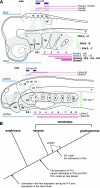
Evolution of the gene expression patterns and the origin of the jaw. (A) Simplified Hox codes in gnathostomes (top) and the lamprey larva (bottom) are summarized. Gnathostome-specific regulatory gene expression domains are shown in bold, and crest cells in grey. In gnathostomes (in this figure, amniotes), distinct sets of Hox transcripts are distributed in a nested pattern in the pharyngeal arches (PAs) with the mandibular arch (PA1) defined by the Hox-free default state and by the expression of Otx cognates. Along the dorsoventral axis of the arches, a Dlx code is established to differentiate the dorsoventral pattern of each PA. In the lamprey, such nested expression of Dlx genes has not been detected. Note that the PA1 is commonly at the state of Hox-free default, and PG2 and PG3 Hox genes have the same rostral boundaries of expression along the PAs between lamprey and gnathostomes, implying the deep origin of the three morphological identities to differentiate PA1, PA2 and the rest of the arches. (B)Evolutionary changes in regulations of Otx expression, Dlx-, and Hox codes have been placed along the phylogenetic tree. Abbreviations: hy, hyoid arch; llp, lower lip; mhb, mid-hindbrain boundary; mn, mandibular process; mx, maxillary process; n, notochord; ot, otic vesicle; r1–5, rhombomeres; ulp, upper lip; vel, velum; 1–8, pharyngeal slits or pouches. Based on Murakami et al. (2004) and Takio et al. (2004).
Fig. 4
Fig. 4
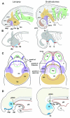
Comparison of mesenchymal developmental patterning. In the lamprey and gnathostome embryonic heads, shared patterns of mesodermal (yellow) and ectomesenchymal (green) distribution can be detected. The endodermal derivatives are coloured pink. The crest cells rostral to the first pharyngeal pouch (pp1) are collectively called the trigeminal crest cells that can be divided into those in the mandibular arch (mandibular crest cells; MC) and the premandibular crest cells (PMC). The posterior region comprising the mandibular crest cells surrounds the premandibular mesoderm (pmm) that arises rostral to the tip of the notochord (n). Different subsets of the trigeminal crest cells are patterned on this shared pattern on mesenchymal distribution, as the oral apparatus in each animal groups arises through different distribution of growth factors such as FGF8 and BMP4 that define the mouth (shown by blue bars). In the lamprey, the upper lip arises from PMC and the lower lip from MC, whereas in gnathostomes both the upper and the lower jaw arise from MC, and the PMC differentiate into the prechordal cranial elements. Blue arrows indicate the position of mouth openings. Abbreviations: Ba1–2, branchial arches; di, diencephalon; e, eye; hm, hyoid mesoderm; ph, pharynx; pp1–3, pharyngeal pouches; pog, preoral gut.
Fig. 5
Fig. 5
Comparisons of oral patterning. Distribution patterns of cephalic crest (A), expression of Fgf8/17 and Bmp2/4 cognates in the ectoderm (B), ventral view of embryonic oral regions (C), and the medial sagittal sections (D) are schematically represented. (A) In the early pharyngula, cephalic crest cells form three distinct cell populations called trigeminal, hyoid (HC) and branchial crest cells (BC). The trigeminal crest cell population can be further subdivided into the mandibular crest cells (mc) and two domains of premandibular crest cells (pmc), based on the topographical relationships with the first pharyngeal pouch (p1), eye (e), premandibular mesoderm (pmm) and the mandibular mesoderm. Note that in the gnathostome embryo the maxillary process (mx) or the upper jaw primordium secondarily grows from the dorsal part of the mandibular arch (arrow; for descriptions see Kuratani & Horigome, 2000; Kuratani et al. 2001). (B) Expressions of Fgf8/17 (blue lines) and Bmp2/4 (red lines) are shown on embryos at similar stages to those in A. (C,D) In the lamprey, nasal placodes and hypophysial placodes develop as a single primordium, the nasohypophysial plate (nhp) that lies rostral to the oral ectoderm (oe). Because of the presence of this plate, the premandibular crest cells (pmc) form the upper lip (ulp) that grows beneath the plate as the floor of the nasohypophysial duct. In the gnathostomes, the hypophysis arises as a part of the oral ectoderm, or Rathke's pouch (Rp), which develops separate from the nasal placodes (np), leaving a median space, which permits the premandibular (pmc) and mandibular crest cells (mc) to grow to form the trabecula and upper jaw, respectively. For induction of the hypophysis, the posterior part of the nasohypophysial plate in the lamprey has to grow posteriorly to reach the hypothalamic anlage (ht). The same topographical relationship is established early in gnathostome development. Abbreviations: lj, lower jaw; llp, lower lip; mo, mouth; n, notochord; ot, otic vesicle; ph, pharynx; vel, velum.
Fig. 6
Fig. 6
Comparisons of equivalent ectomesenchymal regions between the lamprey and gnathostomes. Terminology for the crest cell populations and subpopulations is based on the topographical distribution of the crest cells with respect to the other embryonic structures such as mesoderm and pharyngeal pouches, not in terms of their developmental fates. This division defines the morphological homology of ectomesenchymal portions, which can be applied throughout vertebrates, laid down at the early pharyngula stages (see Figs 4 and 5). Note, however, that different portions of ectomesenchyme are utilized to differentiate into the ‘oral apparatus’ in the lamprey and gnathostomes (shaded). Because of this heterotopic shift in tissue interactions, the ectomesenchymal part with the same name in the lamprey and gnathostomes do not always differentiate into the same skeletal elements (see Kuratani et al. 2001; Shigetani et al. 2002). For the homology of the cartilages called ‘trabeculae’ between the two animal groups, see Kuratani et al. (2004).

References

    1. Barlow AJ, Francis-West PH. Ectopic application of recombinant BMP-2 and BMP-4 can change patterning of developing chick facial primordia. Development. 1997;124:391–398. - PubMed
    1. de Beer GR. On the nature of the trabecula cranii. Q. J. Microsc. Sci. 1931;74:701–731.
    1. de Beer GR. The Development of the Vertebrate Skull. London: Oxford University Press; 1937.
    1. Boorman CJ, Shimeld SM. Cloning and expression of a Pitx homeobox gene from the lamprey, a jawless vertebrate. Dev. Genes Evol. 2002;212:349–353. - PubMed
    1. Carroll RL. Vertebrate Paleontology and Evolution. New York: W.H. Freeman; 1988.

Publication types

LinkOut - more resources