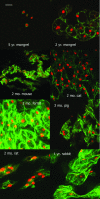
Cytomorphology of notochordal and chondrocytic cells from the nucleus pulposus: a species comparison
- PMID: 15575884
- PMCID: PMC1571361
- DOI: 10.1111/j.0021-8782.2004.00352.x
Cytomorphology of notochordal and chondrocytic cells from the nucleus pulposus: a species comparison
Abstract
The nuclei pulposi of the intervertebral discs (IVDs) contain a mixed population of cell types at various stages of maturation. This tissue is formed either by or with the help of cells from the embryonic notochord, which appear to be replaced during development by a population of chondrocyte-like cells of uncertain origin. However, this transition occurs at widely varying times, depending upon the species--or even breed--of the animal being examined. There is considerable debate among spine researchers as to whether the presence of these residual notochordal cells has a significant impact upon IVD degeneration models, and thus which models may best represent the human condition. The present study examines several different species commonly used in lumbar spine investigations to explore the variability of notochordal cells in the IVD.
Figures


References
-
- Adler JH, Schoenbaum M, Silberberg R. Early onset of disk degeneration and spondylosis in sand rats (Psammomys obesus) Vet. Pathol. 1983;20:13–22. - PubMed
-
- Aguiar DJ, Johnson SL, Oegema TR. Notochordal cells interact with nucleus pulposus cells: regulation of proteoglycan synthesis. Exp. Cell Res. 1999;246:129–137. - PubMed
-
- Anderson DG, Izzo MW, Hall DJ, et al. Comparative gene expression profiling of normal and degenerative discs: analysis of a rabbit annular laceration model. Spine. 2002;27:1291–1296. - PubMed
-
- Ariga K, Miyamoto S, Nakase T, et al. The relationship between apoptosis of endplate chondrocytes and aging and degeneration of the intervertebral disc. Spine. 2001;26:2414–2420. - PubMed
-
- Bell GR. Anatomy of the lumbar spine: developmental to normal adult anatomy. In: Wiesel SW, Weinstein JN, Herkowitz HN, Dvorak J, Bell GR, editors. The Lumbar Spine. Philadelphia, PA: W.B. Saunders Company; 1996. pp. 43–52.
Publication types
MeSH terms
LinkOut - more resources
Full Text Sources

