. 2004 Dec 1;32(21):6251-9.
doi: 10.1093/nar/gkh962. Print 2004.

DmGEN, a novel RAD2 family endo-exonuclease from Drosophila melanogaster

Affiliations

DmGEN, a novel RAD2 family endo-exonuclease from Drosophila melanogaster

Gen Ishikawa et al. Nucleic Acids Res. .

Abstract

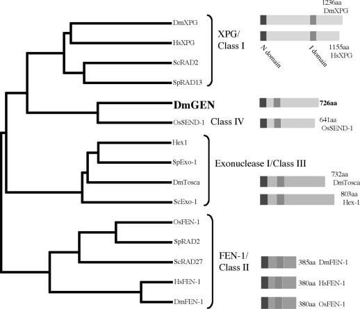
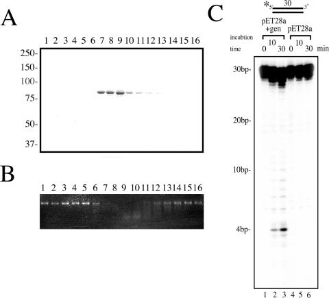
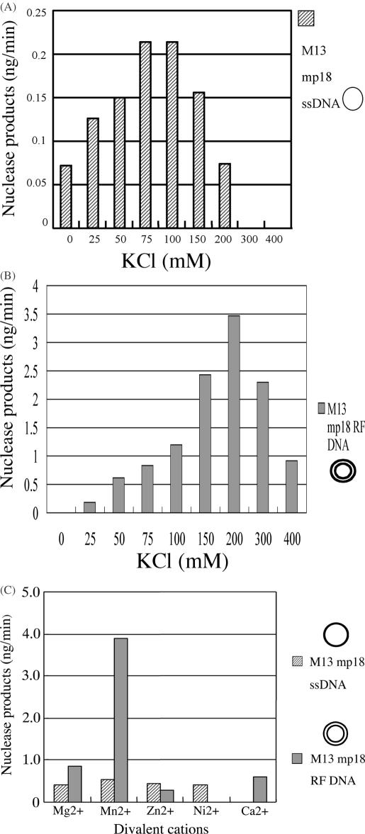
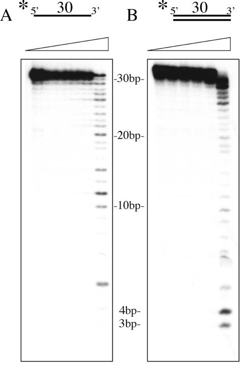
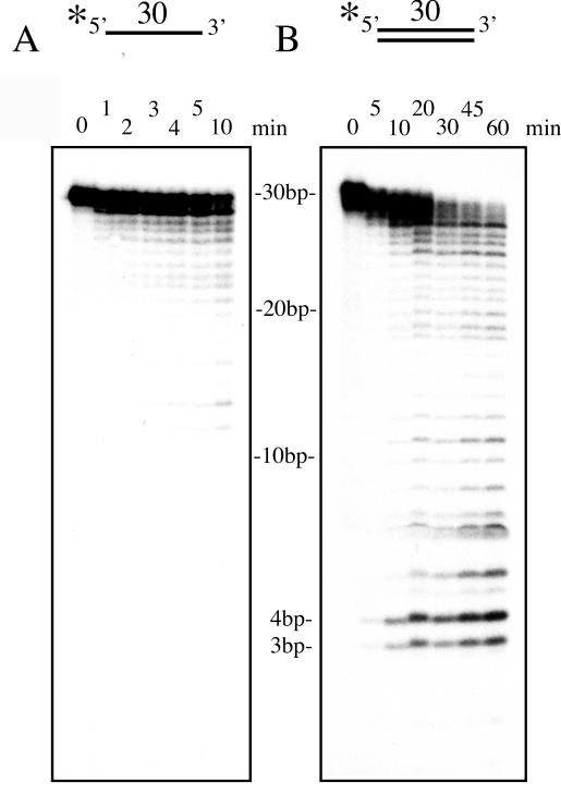
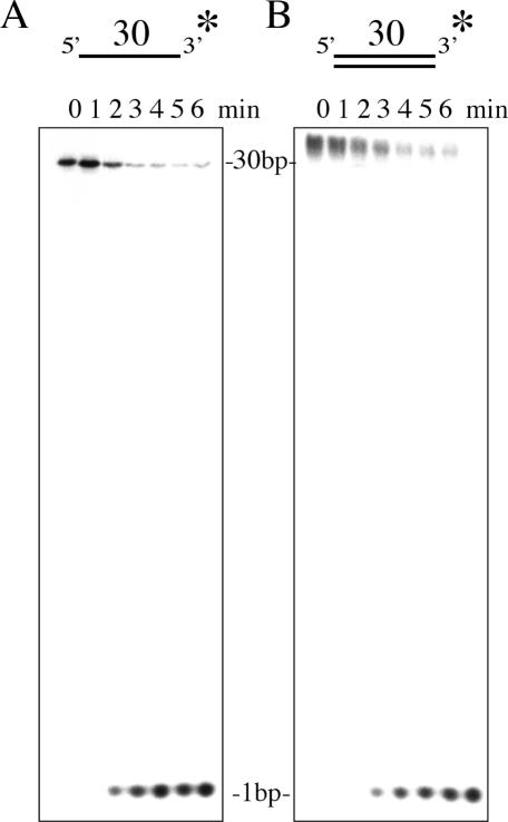
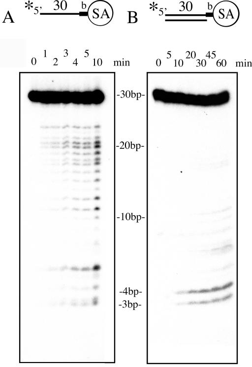
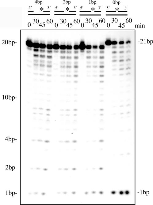
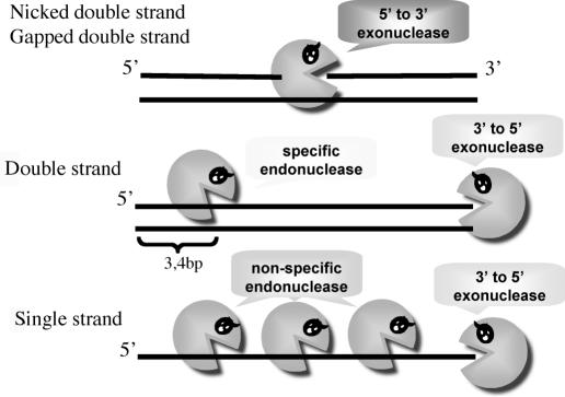
A novel endo-exonuclease, DmGEN (Drosophila Melanogaster XPG-like endonuclease), was identified in D.melanogaster. DmGEN is composed of five exons and four introns, and the open reading frame encodes a predicted product of 726 amino acid residues with a molecular weight of 82.5 kDa and a pI of 5.36. The gene locus on Drosophila polytene chromosomes was detected at 64C9 on the left arm of chromosome 3 as a single site. The encoded protein showed a relatively high degree of sequence homology with the RAD2 nucleases, especially XPG. Although the XPG-N- and XPG-I-domains are highly conserved in sequence, locations of the domains are similar to those of FEN-1 and EXO-1, and the molecular weight of the protein is close to that of EXO-1. In vitro, DmGEN showed endonuclease and 3'-5' exonuclease activities with both single-stranded DNA (ssDNA) and double-stranded DNA (dsDNA), but the endonuclease action with dsDNA was quite specific: 5'-3' exonuclease activity was found to occur with nicked DNA, while dsDNA was endonucleolytically cut at 3-4 bp from the 5' end. Homologs are widely found in mammals and higher plants. The data suggest that DmGEN belongs to a new class of RAD2 nuclease.


Figures

Figure 1
Figure 1
The dendrogram on the left depicts relationships among FEN-1/RAD2 family members. On the right are depicted the protein structures with highly conserved domains (XPG-N domain and XPG-I domain) indicated.
Figure 2
Figure 2
(A) Purification of DmGEN protein. DmGEN protein overexpressed in E.coli was separated by 7.5% SDS–PAGE and stained with Coomassie brilliant blue. (B) Endonuclease activity of each fraction from (A). The substrate was single-strand circular DNA. Incubation was at 37°C for 30 min. (C) Nuclease activity on double-stranded DNA (dsDNA) to confirm expression of the DmGEN protein. The total reaction volume was 20 μl. 5′ end-labeled dsDNA (100 fmol) was incubated with the protein (16.2 ng) extracted from E.coli. Time course experiments were performed. Schematic representations of the substrates are shown. The asterisk indicates the position of the radiolabel. Substrate and cleavage product sizes were as indicated. Lanes 1–3 included extracts from E.coli carrying the pET28a+ vector with the DmGEN gene. Lanes 4–6 included extracts from E.coli with the pET28a+ vector alone. Reaction products were resolved on 20% polyacrylamide/8 M urea gels and visualized using autoradiography.
Figure 3
Figure 3
The basic biochemical properties of the DmGEN protein. (A) Effects of salt concentration on the endonuclease activity of DmGEN protein (32.4 ng) toward ssDNA substrate. Incubation was conducted at 37°C for 60 min with or without DmGEN proteins. (B) Effects of salt concentration on the endonuclease activity of DmGEN protein (32.4 ng) toward dsDNA substrate. Incubation was conducted at 37°C for 10 min with or without DmGEN proteins. (C) Effects of divalent cations on the endonuclease activity of the DmGEN protein (32.4 ng) toward ssDNA and dsDNA substrates. Incubation was conducted at 37°C for 30 min with DmGEN proteins. Samples were analyzed by electrophoresis on 0.7% agarose gels. Amounts of nuclease products were calculated with the aid of a luminescent image analyzer (Fujifilm).
Figure 4
Figure 4
Incubation of different amounts of DmGEN protein in nuclease assays. Ramps indicate increasing amounts of DmGEN in 20 μl of reaction mixture (0, 1.0, 2.0, 4.1, 8.1, 16.2 ng). DmGEN protein was incubated with 100 fmol of ssDNA (A) or dsDNA (B) for 30 min at 37°C.
Figure 5
Figure 5
Nuclease activity of DmGEN protein (16.2 ng) on linear DNA substrates. Time course experiments were performed. The substrates are depicted schematically in each panel. The asterisk indicates the position of the radiolabel. Substrate and cleavage product sizes were as indicated. (A) The substrate was single-strand labeled at the 5′ end. (B) The substrate was double-strand labeled at the 5′ end.
Figure 6
Figure 6
Nuclease activity of DmGEN protein (16.2 ng) on linear DNA substrates. Time course experiments were performed. The substrates are depicted schematically in each panel. The asterisk indicates the position of the radiolabel. Substrate and cleavage product sizes were as indicated. (A) The substrate was single-strand labeled at the 3′ end. (B) The substrate was double-strand labeled at the 3′ end.
Figure 7
Figure 7
Effects of biotin adducts and streptavidin binding to single- and double-stranded substrates on the exonuclease activity of DmGEN protein (16.2 ng). Time course experiments were performed. The substrates are depicted schematically in each panel. The asterisk indicates the position of the radiolabel. Substrate and cleavage product sizes were as indicated. (A) The substrate was single-strand labeled at the 5′ end. (B) The substrate was double-strand labeled at the 5′ end.
Figure 8
Figure 8
Nuclease activity of DmGEN protein (16.2 ng) on different substrates. Time course experiments were performed. Ramps indicate an increase in the reaction time (0, 30, 45 and 60 min). The substrates are depicted schematically in each panel. The asterisk indicates the position of the radiolabel. The sizes of gaps and cleavage products were as indicated.
Figure 9
Figure 9
Summary of cleavage sites.

References

    1. Kimura S., Kai,M., Kobayashi,H., Suzuki,A., Morioka,H., Otsuka,E. and Sakaguchi,K. (1997) A structure-specific endonuclease from cauliflower (Brassica oleracea var. botrytis) inflorescence. Nucleic Acids Res., 25, 4970–4976. - PMC - PubMed
    1. Kimura S., Suzuki,T., Yanagawa,Y., Yamamoto,T., Nakagawa,H., Tanaka,I., Hashimoto,J. and Sakaguchi,K. (2001) Characterization of plant proliferating cell nuclear antigen (PCNA) and flap endonuclease-1 (FEN-1), and their distribution in mitotic and meiotic cell cycles. Plant J., 28, 643–653. - PubMed
    1. Kimura S., Ueda,T., Hatanaka,M., Takenouchi,M., Hashimoto,J. and Sakaguchi,K. (2000) Plant homologue of flap endonuclease-1: molecular cloning, characterization, and evidence of expression in meristematic tissues. Plant Mol. Biol., 42, 415–427. - PubMed
    1. Furukawa T., Kimura,S., Ishibashi,T., Mori,Y., Hashimoto,J. and Sakaguchi,K. (2003) OsSEND-1: a new RAD2 nuclease family member in higher plants. Plant Mol. Biol., 51, 59–70. - PubMed
    1. Calleja F.M., Nivard,M.J. and Eeken,J.C. (2001) Induced mutagenic effects in the nucleotide excision repair deficient Drosophila mutant mus201(D1), expressing a truncated XPG protein. Mutat. Res., 461, 279–288. - PubMed

MeSH terms

Associated data