The length of the reactive center loop modulates the latency transition of plasminogen activator inhibitor-1
- PMID: 15576554
- PMCID: PMC2253313
- DOI: 10.1110/ps.041063705
The length of the reactive center loop modulates the latency transition of plasminogen activator inhibitor-1
Abstract
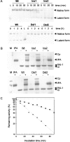
Plasminogen activator inhibitor-1 (PAI-1) belongs to the serine protease inhibitor (serpin) protein family, which has a common tertiary structure consisting of three beta-sheets and several alpha-helices. Despite the similarity of its structure with those of other serpins, PAI-1 is unique in its conformational lability, which allows the conversion of the metastable active form to a more stable latent conformation under physiological conditions. For the conformational conversion to occur, the reactive center loop (RCL) of PAI-1 must be mobilized and inserted into the major beta-sheet, A sheet. In an effort to understand how the structural conversion is regulated in this conformationally labile serpin, we modulated the length of the RCL of PAI-1. We show that releasing the constraint on the RCL by extension of the loop facilitates a conformational transition of PAI-1 to a stable state. Biochemical data strongly suggest that the stabilization of the transformed conformation is owing to the insertion of the RCL into A beta-sheet, as in the known latent form. In contrast, reducing the loop length drastically retards the conformational change. The results clearly show that the constraint on the RCL is a factor that regulates the conformational transition of PAI-1.
Figures


Similar articles
-
Structural factors affecting the choice between latency transition and polymerization in inhibitory serpins.Protein Sci. 2007 May;16(5):833-41. doi: 10.1110/ps.062745807. Epub 2007 Mar 30. Protein Sci. 2007. PMID: 17400919 Free PMC article.
-
Specific interactions of serpins in their native forms attenuate their conformational transitions.Protein Sci. 2007 Aug;16(8):1659-66. doi: 10.1110/ps.072838107. Epub 2007 Jun 28. Protein Sci. 2007. PMID: 17600149 Free PMC article.
-
Conformational changes of the reactive-centre loop and beta-strand 5A accompany temperature-dependent inhibitor-substrate transition of plasminogen-activator inhibitor 1.Eur J Biochem. 1996 Oct 1;241(1):38-46. doi: 10.1111/j.1432-1033.1996.0038t.x. Eur J Biochem. 1996. PMID: 8898886
-
Biochemical properties of plasminogen activator inhibitor-1.Front Biosci (Landmark Ed). 2009 Jan 1;14(4):1337-61. doi: 10.2741/3312. Front Biosci (Landmark Ed). 2009. PMID: 19273134 Review.
-
The molecular basis for anti-proteolytic and non-proteolytic functions of plasminogen activator inhibitor type-1: roles of the reactive centre loop, the shutter region, the flexible joint region and the small serpin fragment.Biol Chem. 2002 Jan;383(1):21-36. doi: 10.1515/BC.2002.003. Biol Chem. 2002. PMID: 11928815 Review.
Cited by
-
Single fluorescence probes along the reactive center loop reveal site-specific changes during the latency transition of PAI-1.Protein Sci. 2016 Feb;25(2):487-98. doi: 10.1002/pro.2839. Epub 2015 Nov 25. Protein Sci. 2016. PMID: 26540464 Free PMC article.
-
Structural factors affecting the choice between latency transition and polymerization in inhibitory serpins.Protein Sci. 2007 May;16(5):833-41. doi: 10.1110/ps.062745807. Epub 2007 Mar 30. Protein Sci. 2007. PMID: 17400919 Free PMC article.
-
Metals affect the structure and activity of human plasminogen activator inhibitor-1. I. Modulation of stability and protease inhibition.Protein Sci. 2011 Feb;20(2):353-65. doi: 10.1002/pro.568. Protein Sci. 2011. PMID: 21280127 Free PMC article.
-
Specific interactions of serpins in their native forms attenuate their conformational transitions.Protein Sci. 2007 Aug;16(8):1659-66. doi: 10.1110/ps.072838107. Epub 2007 Jun 28. Protein Sci. 2007. PMID: 17600149 Free PMC article.
References
-
- Anfinsen, C.B. 1973. Principles that govern the folding of protein chains. Science 181 223–230. - PubMed
-
- Carrell, R.W., Evans, D.L., and Stein, P.E. 1991. Mobile reactive centre of serpins and the control of thrombosis. Nature 353 576–578. - PubMed
-
- Carrell, R.W., Stein, P.E., Fermi, G., and Wardell, M.R. 1994. Biological implication of a 3 Å structure of dimeric antithrombin. Structure 2 257–270. - PubMed
-
- Carrell, R.W., Huntington, J.A., Mushunje, A., and Zhou, A. 2001. The conformational basis of thrombosis. Thromb. Haemost. 86 14–22. - PubMed
Publication types
MeSH terms
Substances
LinkOut - more resources
Full Text Sources
Miscellaneous

