. 2004 Dec 2;32(21):6378-87.
doi: 10.1093/nar/gkh980. Print 2004.

PriA helicase and SSB interact physically and functionally

Affiliations

PriA helicase and SSB interact physically and functionally

Chris J Cadman et al. Nucleic Acids Res. .

Abstract

PriA helicase is the major DNA replication restart initiator in Escherichia coli and acts to reload the replicative helicase DnaB back onto the chromosome at repaired replication forks and D-loops formed by recombination. We have discovered that PriA-catalysed unwinding of branched DNA substrates is stimulated specifically by contact with the single-strand DNA binding protein of E.coli, SSB. This stimulation requires binding of SSB to the initial DNA substrate and is effected via a physical interaction between PriA and the C-terminus of SSB. Stimulation of PriA by the SSB C-terminus may act to ensure that efficient PriA-catalysed reloading of DnaB occurs only onto the lagging strand template of repaired forks and D-loops. Correlation between the DNA repair and recombination defects of strains harbouring an SSB C-terminal mutation with inhibition of this SSB-PriA interaction in vitro suggests that SSB plays a critical role in facilitating PriA-directed replication restart. Taken together with previous data, these findings indicate that protein-protein interactions involving SSB may coordinate replication fork reloading from start to finish.


Figures
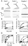
Figure 1
Figure 1
Effect of wild-type SSB on PriA helicase function. (A) Unwinding reactions were performed with 0.2 nM PriA as indicated in the presence of 0, 0.02, 0.05, 0.2, 2 and 10 nM SSB tetramers (lanes 2–7). Incubation with 10 nM SSB tetramers in the absence of PriA was performed (lane 8) to ensure any dissociation was not due to duplex melting by SSB. DNA substrates are depicted above each gel whilst products of unwinding are shown on the left. Positions of 32P labels are shown as circles, arrows mark the 3′ ends of DNA strands and numbers indicate the length in nucleotides of each arm. The 50mer and 70mer oligonucleotides correspond to the leading and lagging strands, respectively, at a replication fork. (B) Quantification of the degree of unwinding in the presence of increasing concentrations of SSB. Closed circles, fork 1; open triangles, fork 2; closed triangles, duplex 1. (C) Rates of unwinding of fork 2 by 0.2 nM PriA in the absence of SSB (closed triangles) and the presence of 2 nM SSB tetramers (open triangles). (D) Binding of forks 1 and 2 and duplex 1 by PriA as monitored by bandshift assays. Symbols are as described in B. (E) Binding of forks 1 and 2 and duplex 1 by SSB. Symbols are as described in (B).
Figure 2
Figure 2
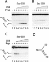
Specificity of SSB-directed stimulation of PriA. (A) Unwinding of fork 2 by 0.2 nM PriA in the presence of 0, 0.05, 0.2, 0.5, 2, 10 and 50 nM E.coli SSB tetramers (lanes 2–8). Lane 9 contains 50 nM SSB tetramers but no PriA. (B) Unwinding of fork 2 by 0.2 nM PriA in the presence of 0, 0.2, 0.8, 2, 8, 40 and 200 nM S.solfataricus SSB monomers (lanes 2–8). Lane 9 contains 200 nM S.solfataricus SSB monomers but no PriA. Concentrations of S.solfataricus SSB monomers were chosen so that equivalent numbers of E.coli and S.solfataricus SSB polypeptides were employed in (A) and (B). (C) Unwinding of fork 2 by 20 nM Rep in the presence of 0, 0.05, 0.2, 0.5, 2, 10 and 50 nM E.coli SSB tetramers (lanes 2–8). Lane 9 contains 50 nM SSB tetramers but no Rep. (D) Structure of fork 2 used in (A), (B) and (C).
Figure 3
Figure 3
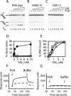
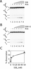
Stimulation of PriA helicase requires physical contact with a wild-type SSB C-terminus. (A) Unwinding of fork 2 by 0.5 nM PriA in the presence of 0, 0.1, 0.2, 0.5, 2 and 10 nM wild-type E.coli SSB tetramers (lanes 2–7). Lane 8 contains 10 nM SSB tetramers but no PriA. (B) Effect of SSBΔC10 on unwinding of fork 2 by PriA. Protein concentrations are as in (A). (C) Effect of SSB113 on unwinding of fork 2 by PriA. Protein concentrations are as in (A). (D) Extent of PriA-catalysed unwinding of fork 2 as a function of SSB concentration. Closed circles, wild-type SSB; open triangles, SSBΔC10; open squares, SSB113. (E) Binding of fork 2 by wild-type and mutant SSBs as monitored by bandshift assays. Symbols are as described in (D). (F) Binding of PriA to a 15mer peptide corresponding to the 15 C-terminal residues of wild-type SSB, as measured by surface plasmon resonance. Times of injection of 200 and 1000 nM PriA (dashed and filled lines, respectively), and of buffer lacking PriA, are indicated by arrows. (G) Binding of PriA to a 15mer peptide corresponding to the 15 C-terminal residues of SSB113. Line designations are as for (F).
Figure 4
Figure 4
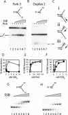
Stimulation of PriA helicase by SSB correlates with binding of SSB to ssDNA within the initial DNA substrate. Unwinding of fork 3 (A) and duplex 2 (B) by 0.2 nM PriA in the presence of 0, 0.05, 0.2, 2 and 10 nM wild-type SSB tetramers (lanes 2–6, respectively). Lane 7 contains 10 nM SSB tetramers but no PriA. (C) Potential translocation activities of PriA, depicted as a shaded triangle, on fork 3 (i and ii) and on duplex 2 (iii). (D) Extent of PriA-catalysed unwinding of fork 3 (open squares) and duplex 2 (closed squares) as a function of SSB concentration. (E) Binding of PriA to fork 3 (open squares) and duplex 2 (closed squares) as measured by bandshift assays. (F) Binding of SSB to fork 3 (open squares) and duplex 2 (closed squares) as measured by bandshift assays. (G) Bandshift assay of wild-type SSB binding to fork 3. Concentrations of SSB tetramers were 0, 0.1, 0.2, 0.4, 0.8, 1.6, 3.2 and 50 nM. (H) Bandshift assay of SSB binding to duplex 2. Concentrations of SSB are as described in (G).
Figure 5
Figure 5
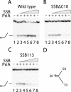
Stimulation of PriA by SSB binding to ssDNA within the initial substrate requires a wild-type SSB C-terminus. Unwinding of fork 3 by 0.5 nM PriA in the presence of 0, 0.1, 0.2, 0.5, 2 and 10 nM tetramers of wild-type SSB (A), SSBΔC10 (B) and SSB113 (C) (lanes 2–7). Lane 8 contains 10 nM SSB tetramers but lacks PriA. (D) Structure of fork 3 used in (A), (B) and (C).
Figure 6
Figure 6
SSB can also stimulate PriA by trapping intermediates of unwinding. (A) Unwinding of fork 1 by 0.2 nM PriA in the presence of 0, 0.05, 0.2, 2, 10 and 50 nM wild-type SSB tetramers (lanes 2–7). Lane 8 contains 50 nM SSB tetramers but no PriA. (B) As for (A) except SSB113 was used in place of wild-type SSB. (C) Levels of stimulation of PriA-catalysed unwinding of fork 1 by wild-type SSB (closed circles) and SSB113 (open squares).

References

    1. Lindahl T. (1996) The Croonian Lecture, 1996: endogenous damage to DNA. Philos. Trans. R. Soc. Lond. B Biol. Sci., 351, 1529–1538. - PubMed
    1. Vilette D., Ehrlich,S.D. and Michel,B. (1995) Transcription-induced deletions in Escherichia coli plasmids. Mol. Microbiol., 17, 493–504. - PubMed
    1. McGlynn P. and Lloyd,R.G. (2000) Modulation of RNA polymerase by (p)ppGpp reveals a RecG-dependent mechanism for replication fork progression. Cell, 101, 35–45. - PubMed
    1. Huertas P. and Aguilera,A. (2003) Cotranscriptionally formed DNA:RNA hybrids mediate transcription elongation impairment and transcription-associated recombination. Mol. Cell, 12, 711–721. - PubMed
    1. Cox M.M., Goodman,M.F., Kreuzer,K.N., Sherratt,D.J., Sandler,S.J. and Marians,K.J. (2000) The importance of repairing stalled replication forks. Nature, 404, 37–41. - PubMed

Publication types

MeSH terms