

Conserved repeat motifs and glucan binding by glucansucrases of oral streptococci and Leuconostoc mesenteroides
- PMID: 15576779
- PMCID: PMC532428
- DOI: 10.1128/JB.186.24.8301-8308.2004
Conserved repeat motifs and glucan binding by glucansucrases of oral streptococci and Leuconostoc mesenteroides
Abstract
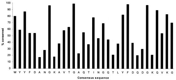
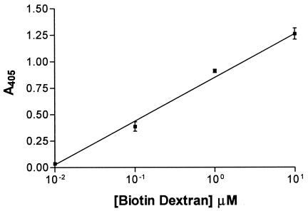
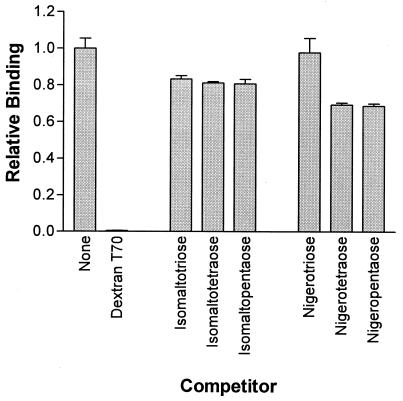
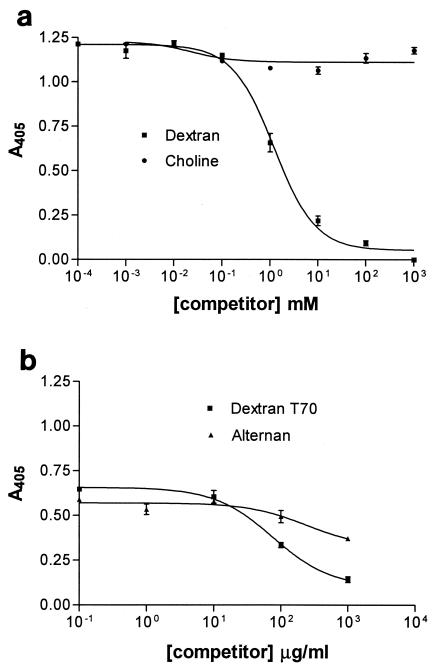
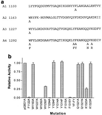
Glucansucrases of oral streptococci and Leuconostoc mesenteroides have a common pattern of structural organization and characteristically contain a domain with a series of tandem amino acid repeats in which certain residues are highly conserved, particularly aromatic amino acids and glycine. In some glucosyltransferases (GTFs) the repeat region has been identified as a glucan binding domain (GBD). Such GBDs are also found in several glucan binding proteins (GBP) of oral streptococci that do not have glucansucrase activity. Alignment of the amino acid sequences of 20 glucansucrases and GBP showed the widespread conservation of the 33-residue A repeat first identified in GtfI of Streptococcus downei. Site-directed mutagenesis of individual highly conserved residues in recombinant GBD of GtfI demonstrated the importance of the first tryptophan and the tyrosine-phenylalanine pair in the binding of dextran, as well as the essential contribution of a basic residue (arginine or lysine). A microplate binding assay was developed to measure the binding affinity of recombinant GBDs. GBD of GtfI was shown to be capable of binding glucans with predominantly alpha-1,3 or alpha-1,6 links, as well as alternating alpha-1,3 and alpha-1,6 links (alternan). Western blot experiments using biotinylated dextran or alternan as probes demonstrated a difference between the binding of streptococcal GTF and GBP and that of Leuconostoc glucansucrases. Experimental data and bioinformatics analysis showed that the A repeat motif is distinct from the 20-residue CW motif, which also has conserved aromatic amino acids and glycine and which occurs in the choline-binding proteins of Streptococcus pneumoniae and other organisms.
Figures
Similar articles
-
Location of repeat elements in glucansucrases of Leuconostoc and Streptococcus species.FEMS Microbiol Lett. 2000 Nov 1;192(1):53-7. doi: 10.1111/j.1574-6968.2000.tb09358.x. FEMS Microbiol Lett. 2000. PMID: 11040428
-
Mutagenesis of asp-569 of glucosyltransferase I glucansucrase modulates glucan and oligosaccharide synthesis.Appl Environ Microbiol. 2000 May;66(5):1923-7. doi: 10.1128/AEM.66.5.1923-1927.2000. Appl Environ Microbiol. 2000. PMID: 10788361 Free PMC article.
-
Definition of a fundamental repeating unit in streptococcal glucosyltransferase glucan-binding regions and related sequences.J Dent Res. 1994 Jun;73(6):1133-41. doi: 10.1177/00220345940730060201. J Dent Res. 1994. PMID: 8046101
-
Glucansucrases: mechanism of action and structure-function relationships.FEMS Microbiol Rev. 1999 Apr;23(2):131-51. doi: 10.1111/j.1574-6976.1999.tb00394.x. FEMS Microbiol Rev. 1999. PMID: 10234842 Review.
-
Glycosyltransferase-mediated Sweet Modification in Oral Streptococci.J Dent Res. 2015 May;94(5):659-65. doi: 10.1177/0022034515574865. Epub 2015 Mar 9. J Dent Res. 2015. PMID: 25755271 Free PMC article. Review.
Cited by
-
LiaS regulates virulence factor expression in Streptococcus mutans.Infect Immun. 2008 Jul;76(7):3093-9. doi: 10.1128/IAI.01627-07. Epub 2008 May 5. Infect Immun. 2008. PMID: 18458070 Free PMC article.
-
Unprecedented Diversity of the Glycoside Hydrolase Family 70: A Comprehensive Analysis of Sequence, Structure, and Function.J Agric Food Chem. 2024 Jul 31;72(30):16911-16929. doi: 10.1021/acs.jafc.4c04807. Epub 2024 Jul 18. J Agric Food Chem. 2024. PMID: 39025827 Free PMC article.
-
Polyphenol Utilization Proteins in the Human Gut Microbiome.Appl Environ Microbiol. 2022 Feb 8;88(3):e0185121. doi: 10.1128/AEM.01851-21. Epub 2021 Dec 1. Appl Environ Microbiol. 2022. PMID: 34851722 Free PMC article.
-
Inhibition of Streptococcus mutans biofilm formation by Streptococcus salivarius FruA.Appl Environ Microbiol. 2011 Mar;77(5):1572-80. doi: 10.1128/AEM.02066-10. Epub 2011 Jan 14. Appl Environ Microbiol. 2011. PMID: 21239559 Free PMC article.
-
Functional role and folding properties of the glucan-binding domain of oral bacterial glucansucrase.FEBS Lett. 2025 Aug;599(16):2388-2402. doi: 10.1002/1873-3468.70128. Epub 2025 Aug 2. FEBS Lett. 2025. PMID: 40751592 Free PMC article.
References
-
- Bailey, T. L., and C. Elkan. 1994. Fitting a mixture model by expectation maximization to discover motifs in biopolymers. Proc. Int. Conf. Intell. Syst. Mol. Biol. 2:28-36. - PubMed
-
- Bailey, T. L., and M. Gribskov. 1998. Combining evidence using p-values: application to sequence homology searches. Bioinformatics 14:48-54. - PubMed
-
- Banas, J. A., and M. M. Vickerman. 2003. Glucan-binding proteins of the oral streptococci. Crit. Rev. Oral Biol. Med. 14:89-99. - PubMed
Publication types
MeSH terms
Substances
Grants and funding
LinkOut - more resources
Full Text Sources
Other Literature Sources
Research Materials
