. 2004 Dec;114(11):1550-63.
doi: 10.1172/JCI21454.

Cardiac adenoviral S100A1 gene delivery rescues failing myocardium

Affiliations

Cardiac adenoviral S100A1 gene delivery rescues failing myocardium

Patrick Most et al. J Clin Invest. 2004 Dec.

Abstract

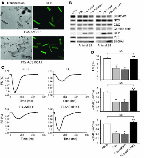
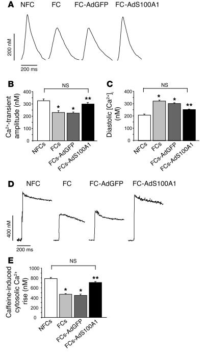
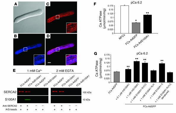
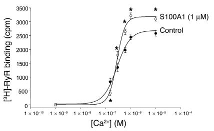
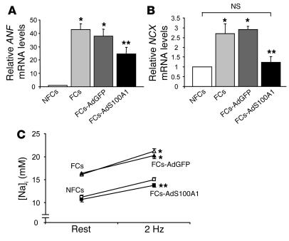
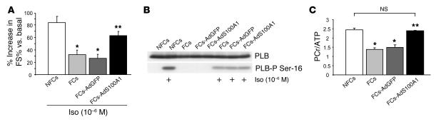
Cardiac-restricted overexpression of the Ca2+-binding protein S100A1 has been shown to lead to increased myocardial contractile performance in vitro and in vivo. Since decreased cardiac expression of S100A1 is a characteristic of heart failure, we tested the hypothesis that S100A1 gene transfer could restore contractile function of failing myocardium. Adenoviral S100A1 gene delivery normalized S100A1 protein expression in a postinfarction rat heart failure model and reversed contractile dysfunction of failing myocardium in vivo and in vitro. S100A1 gene transfer to failing cardiomyocytes restored diminished intracellular Ca2+ transients and sarcoplasmic reticulum (SR) Ca2+ load mechanistically due to increased SR Ca2+ uptake and reduced SR Ca2+ leak. Moreover, S100A1 gene transfer decreased elevated intracellular Na+ concentrations to levels detected in nonfailing cardiomyocytes, reversed reactivated fetal gene expression, and restored energy supply in failing cardiomyocytes. Intracoronary adenovirus-mediated S100A1 gene delivery in vivo to the postinfarcted failing rat heart normalized myocardial contractile function and Ca2+ handling, which provided support in a physiological context for results found in myocytes. Thus, the present study demonstrates that restoration of S100A1 protein levels in failing myocardium by gene transfer may be a novel therapeutic strategy for the treatment of heart failure.


Figures

Figure 1
Figure 1
Postinfarct heart failure model. (A) Representative TTC-stained cross-sections of a sham-operated (Sham-OP, left) and a cryoinfarcted (Cryo-MI, right) rat heart 6 hours after surgery. Transmural cryoinfarcted myocardium emerges as brown tissue with a gray-white border zone (right). Scale bar: 5 mm. (B) Representative mid-ventricular cross sections of a sham-operated (left) and cryoinfarcted (right) rat heart 12 weeks after surgery. Scale bar: 1 mm. (C) Representative images of a freshly isolated LV cardiomyocyte from a nonfailing sham-operated (NFC, left) and cryoinfarcted failing heart (FC, right) 12 weeks after surgery. Note the marked increase in end-diastolic length in the FC. Scale bar: 25 μm.
Figure 2
Figure 2
Reactivated fetal gene expression and abnormal abundance of Ca2+-regulatory proteins in failing myocardium. (A) Reactivated fetal gene expression (ANF, NCX, and α-sk-actin) in rat FCs versus NFCs from sham-OP hearts (n = 6; *P < 0.01, FCs vs. NFCs). Results were obtained from 5 different hearts in each group 12 weeks after surgery. (B) Abnormal protein expression in failing cryoinfarcted rat hearts. Left: Representative results of Western blots for NCX, SERCA2, CSQ, S100A1, and PLB from pooled fractions of FCs (n = 5) and NFCs (n = 5) from cryoinfarcted and sham-OP rat hearts. Results were obtained 12 weeks after surgery. Right: Average fold change in protein expression in failing cryoinfarcted hearts relative to the sham-OP group (n = 7; *P < 0.01, FCs vs. NFCs). Data are given as mean ± SEM.
Figure 3
Figure 3
S100A1-mediated rescue of contractile dysfunction in vitro. (A) Efficiency of adenovirus-mediated gene transfer in FCs. Representative transmission (left) and GFP emission images (right) from FCs 24 hours after adenoviral infection. Upper left: FCs-AdGFP transmission; upper right: FCs-AdGFP, 510-nm emission; lower left: FCs-AdS100A1 transmission; lower right: FCs-AdS100A1, 510-nm emission. Scale bar: 100 μm. (B) Restoration of S100A1 protein levels in FCs 24 hours after S100A1 gene transfer. Representative results of Western blots for SERCA2, NCX, CSQ, cardiac actin, GFP, PLB, and S100A1 from homogenates both of untreated NFCs and FCs and AdGFP- and AdS100A1-transfected FCs. Note that GFP is only expressed in adenovirus-treated failing cells. Data are shown from 2 different representative preparations (animals 82 and 93). (C) Rescue of contractile function in FCs after adenoviral S100A1 gene transfer. Original tracings of FS (shown as downward deflection) from a representative NFC, FC, FC-AdGFP, and FC-AdS100A1. (D) Normalization of FS (upper panel), rate of cellular shortening (_dl/dt; μm/ms; middle panel), and rate of cellular relengthening (+dl/dt; μm/ms; lower panel) in FCs after S100A1 gene addition (n = 40 cells from 4 different preparations in each group; *P < 0.01 compared with NFCs; **P < 0.01 compared with FCs and FCs-AdGFP); P = NS, NFCs vs. FCs-AdS100A1. Data are presented as mean ± SEM.
Figure 4
Figure 4
Normalization of Ca2+ transients and SR Ca2+ load after S100A1 gene delivery in FCs. (A) Original tracings of Ca2+ transients (shown as upward deflection) from a representative NFC, FC, FC-AdGFP, and FC-AdS100A1. (B) Normalization of Ca2+-transient amplitude (nM) and (C) significant decrease in diastolic [Ca2+] (nM) in failing cells after AdS100A1 treatment (n = 40 cells from 4 different preparations in each group). (D) Original tracings of cytosolic Ca2+ rise in response to acute caffeine application (10 mM) in the presence of Ni2+ (5 mM) (shown as upward deflection) from a representative NFC, FC, FC-AdGFP, and FC-AdS100A1. (E) Normalization of SR Ca2+ load estimated from the caffeine-induced cytosolic Ca2+ rise (nM) in FCs after AdS100A1 gene transfer (n = 40 cells from 4 different preparations in each group). *P < 0.01 compared with NFCs; **P < 0.01 compared with FCs and FCs-AdGFP; P = NS, NFCs vs. FCs-AdS100A1. Data are presented as mean ± SEM.
Figure 5
Figure 5
S100A1 interacts with SERCA2 and increases activity of the SR Ca2+ pump in failing myocardium. (A) Nomarski image of an FC-AdS100A1. Immunolabeling of (B) S100A1 (blue) and (C) SERCA2 (red) in the same cell. (D) Overlay of B and C depicts colocalization of SERCA2 and S100A1 (violet). Scale bar: 20 μm. Inset magnification, ×3. (E) Ca2+-dependent coimmunoprecipitation of SERCA2 (red) and S100A1 (green). Samples were immunoprecipated with anti-SERCA2 antibody and costained for S100A1. Control experiments were carried out with A/G-Sepharose beads (A/G-beads) only. (F) Enhancement of Ca2+-dependent Ca2+-ATPase activity in homogenates of FCs-AdS100A1. (G) Coincubation of FC-AdGFP homogenates with human recombinant S100A1 protein (rhS100A1) and S100A1 peptides enhances SERCA2 activity. Experiments were carried out at pCa 6.2 (n = 6). *P < 0.01, NFCs vs. FCs; **P < 0.01, FCs-AdS100A1 and FCs-AdGFP + S100A1 protein or peptides vs. FCs-AdGFP. Pooled cardiomyocyte samples were obtained from 4 different preparations in each group. Data are presented as mean ± SEM.
Figure 6
Figure 6
S100A1 interacts with SERCA2 and increases activity of the SR Ca2+ pump in COS cells. (A) Representative Western blots given for adenovirally expressed S100A1 and SERCA2 in COS cells. β-Actin staining served as loading control. (B) Left: Enhanced Ca2+-dependent ATPase activity in SERCA2-expressing COS cells by coexpressed S100A1 protein. Expression of S100A1 alone did not alter Ca2+-dependent ATPase activity in COS cells. Note that addition of anti-S100A1 antibody abrogated the S100A1-mediated increase in Ca2+-dependent ATPase activity. Right: Increased Ca2+-dependent ATPase activity in SERCA2-expressing COS cells following application of human recombinant S100A1 protein (rh-S100A1, 1 μM). Application of anti-S100A1 antibody (10μl) abrogated the S100A1-mediated enhancement of Ca2+-dependent ATPase activity. Application of the SERCA2 inhibitor thapsigargin (10_6 M) abolished Ca2+-dependent ATPase activity in AdSERCA2-infected COS cells. Experiments were carried out at pCa 6.2 (n = 3). *P < 0.01 vs. AdSERCA2; **P < 0.01 vs. AdS100A1/AdSERCA2. Data are presented as mean ± SEM. (C) Ca2+-dependent coimmunoprecipitation of SERCA2 (red) and S100A1 (green). Samples were immunoprecipated with anti-S100A1 antibody and costained for SERCA2. Control experiments were carried out with an anti-S100A1 antibody preincubated with a blocking peptide.
Figure 7
Figure 7
S100A1 interacts with RyR2 and reduces the SR Ca2+ leak in failing myocardium. (A) Nomarski image of an NFC-AdS100A1. Immunolabeling of (B) S100A1 (blue) and (C) RyR2 (red) in the same cell. (D) Overlay of B and C depicts colocalization of S100A1 and RyR2 (violet). Scale bar: 20 μm. Inset magnification, ×3. (E) Ca2+-dependent coimmunoprecipitation of RyR2 (green) and S100A1 (red). Control experiments were carried out with A/G-Sepharose beads only. (F and G) Typical tracings of the time course (F) and averaged values (%) (G) of the SR Ca2+ leak in nonfailing and failing myocardium and AdGFP- and AdS100A1-transfected failing myocardium. Arrow indicates addition of thapsigargin (+TG; 1 μM) (n = 4). *P < 0.01, NF vs. F and F-AdGFP; **P < 0.01, F-AdS100A1 vs. F and F-AdGFP; P = NS, NF vs. F-AdS100A1. Data are presented as mean ± SEM.
Figure 8
Figure 8
S100A1 modulates RyR2 activity in biphasic manner. Control SR vesicles show a Ca2+-dependent increase of [3H]-RyR binding in the presence of 0.5 mM Mg2+ and 10 mM caffeine. Addition of 1 μM S100A1 protein decreased [3H]-RyR at 150 nM free Ca2+ concentrations, while greater than approximately 300 nM free Ca2+ S100A1 increased [3H]-RyR binding to the cardiac SR Ca2+-release channel. Data are presented as mean ± SEM and expressed as cpm. Experiments (n = 3) were carried out in triplicate. *P < 0.05 vs. control.
Figure 9
Figure 9
Effect of adenovirally expressed S100A1 on fetal gene expression and Na+-handling in FCs. (A) Average relative mRNA expression for ANF (upper panel) and (B) NCX (lower panel). 18S rRNA signals were used for normalization. n = 6. (C) Normalization of [Na+]i in FCs following S100A1 gene transfer (n = 40 cells from 4 different preparations; FCs [open triangles], FCs-AdGFP [filled triangles], NFCs [open squares], FCs-AdS100A1 [filled squares]). *P < 0.01 compared with NFCs; **P < 0.01 compared with FCs and FCs-AdGFP; P = NS, NFCs vs. FCs-AdS100A1. Data are presented as mean ± SEM.
Figure 10
Figure 10
Adenoviral S100A1 gene delivery improves contractile reserve and normalizes energy supply in FCs. (A) Improved β-adrenergic stimulated contractility due to S100A1 gene addition in FCs (n = 30 cells from 3 different preparations). (B) Representative Western blots for total PLB (upper panel) and phosphorylated PLB (PLB-P Ser-16, lower panel) levels in NFCs, FCs, FCs-AdGFP, and FCs-AdS100A1_infected FCs under basal conditions and in response to isoproterenol (Iso, 10_6 M). (C) Normalization of PCr/ATP ratio in FCs following S100A1 gene addition. Average results for PCr/ATP ratio of each experimental group. n = 4; *P < 0.01 compared with NFCs; **P < 0.01 compared with FCs and FCs-AdGFP; P = NS, NFCs vs. FCs-AdS100A1. Data are presented as mean ± SEM.
Figure 11
Figure 11
Intracoronary adenoviral S100A1 gene delivery rescues contractile dysfunction in vivo. (A_F) Representative corresponding Nomarski and GFP-fluorescence images from midventricular cryosections of nonfailing (A and B) and AdS100A1- (C and D) and AdGFP-treated (E and F) failing myocardium. Magnification, ×10. (G) Cardiac S100A1 gene transfer reconstitutes S100A1 protein levels in failing myocardium in vivo. Representative Western blot of S100A1, CSQ and GFP expression in sham-OP nonfailing, saline-treated failing, and adenovirus-treated (AdGFP/AdS100A1) failing myocardium. (H_L) Restoration of basal cardiac contractile performance after AdS100A1 gene transfer in vivo. (M_O) Preserved gain-in-function of AdS100A1-treated failing hearts in vivo after isoproterenol stimulation. Saline- and AdGFP-treated failing hearts displayed no significant difference in functional parameters and were combined to heart failure control group (HF-control; n = 14). Data were obtained in isoflurane-anesthetized animals 7 days after intracoronary gene transfer or saline injection. Sham-OP; n = 7, HF-AdS100A1; n = 7. *P < 0.05 HF-AdS100A1 vs. HF control. BW, body weight.
Figure 12
Figure 12
Intracoronary adenoviral S100A1 gene delivery reverses fetal gene expression and aberrant protein expression in failing myocardium in vivo. (A) Reversed fetal gene expression after S100A1 gene delivery in failing myocardium in vivo (*P < 0.05 HF-AdS100A1 vs. HF-control; n = 4; samples were obtained from 3 different animals in each group). (B) Impact of S100A1 gene addition on SERCA2, PLB, and NCX protein amount in failing myocardium in vivo compared with saline-treated and AdGFP-treated failing myocardium (n = 4; samples were obtained from 3 different animals in each group). Data are presented as mean ±SEM.

Similar articles

Cited by

References

    1. American Heart Association. 2003. Heart disease and stroke statistics — 2004 update. American Heart Association. Dallas, Texas, USA. http://www.americanheart.org/presenter.jhtml?identifier=1928.
    1. Marks AR. Calcium and the heart: a question of life and death. J. Clin. Invest. 2003;111:597–600. doi:10.1172/JCI200318067. - PMC - PubMed
    1. Hasenfuss G. Alterations of calcium-regulatory proteins in heart failure. Cardiovasc. Res. 1998;37:279–289. - PubMed
    1. Donato R. Intracellular and extracellular roles of S100 proteins. Microsc. Res. Tech. 2003;60:540–551. - PubMed
    1. Remppis A, et al. Altered expression of the Ca(2+)-binding protein S100A1 in human cardiomyopathy. . Biochim. Biophys. Acta. 1996; 1313:253–257. - PubMed

Publication types

MeSH terms