. 2004 Dec;114(11):1593-602.
doi: 10.1172/JCI21345.

Specific NEMO mutations impair CD40-mediated c-Rel activation and B cell terminal differentiation

Affiliations

Specific NEMO mutations impair CD40-mediated c-Rel activation and B cell terminal differentiation

Ashish Jain et al. J Clin Invest. 2004 Dec.

Abstract

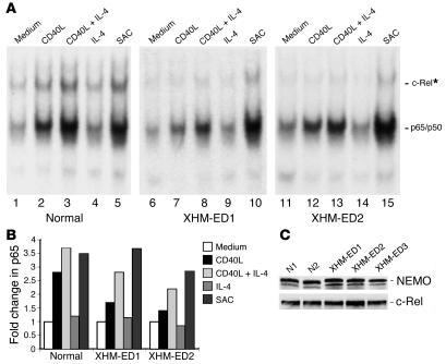
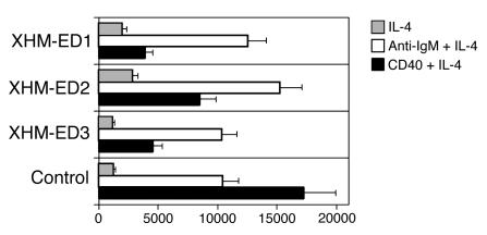
Hypomorphic mutations in the zinc finger domain of NF-kappaB essential modulator (NEMO) cause X-linked hyper-IgM syndrome with ectodermal dysplasia (XHM-ED). Here we report that patient B cells are characterized by an absence of Ig somatic hypermutation (SHM) and defective class switch recombination (CSR) despite normal induction of activation-induced cytidine deaminase (AID) and Iepsilon-Cepsilon transcripts. This indicates that AID expression alone is insufficient to support neutralizing antibody responses. Furthermore, we show that patient B cells stimulated with CD40 ligand are impaired in both p65 and c-Rel activation, and whereas addition of IL-4 can enhance p65 activity, c-Rel activity remains deficient. This suggests that these NF-kappaB components have different activation requirements and that IL-4 can augment some but not all NEMO-dependent NF-kappaB signaling. Finally, using microarray analysis of patient B cells we identified downstream effects of impaired NF-kappaB activation and candidate factors that may be necessary for CSR and SHM in B cells.


Figures

Figure 1
Figure 1
IL-4 synergizes with CD40L in activating p65 binding but not c-Rel binding in XHM-ED B cells. (A) DNA binding activity of the NF-κB complexes. CD19+ B cells were isolated from 2 unrelated patients with the C417R mutation and a normal control. Cells were stimulated in the presence of cycloheximide with CD40L, CD40L + IL-4, IL-4 alone, or Staphylococcus aureus Cowan’s strain I (SAC) for 2 hours. Cellular extracts were prepared and analyzed by electrophoretic mobility shift assay for NF-κB binding activity. *For supershift, all samples were incubated with anti_c-Rel antibody prior to incubation with the labeled probe. One representative experiment of 3 is shown. (B) Quantification of p65 activity. The band shifts for p65 shown in A were quantified and are represented as fold change in comparison to unstimulated cells (set at 1.0). In contrast to the normal control, both XHM-ED1 and XHM-ED2 showed impaired p65 activation in response to CD40 stimulation alone and were enhanced with IL-4 costimulation. (C) NEMO and c-Rel protein levels in B cells. Cytoplasmic extracts were prepared from purified B cells from 2 normal controls and 3 patients with XHM-ED and subjected to immunoblotting. Blots were probed with anti-NEMO or anti_c-Rel antibody.
Figure 2
Figure 2
IL-4 “rescues” the transcriptional regulation of many genes in B cells of XHM-ED patients. B cells from XHM and XHM-ED patients were stimulated with CD40L alone or CD40L + IL-4 for 6 hours or 24 hours. Each row represents the ratio of expression in stimulated vs. unstimulated cells for each gene. Each column represents the data from an independent stimulation experiment. A color bar shows the magnitude of gene expression changes as a ratio of expression: significantly induced (red), unchanged (black), or repressed (green). (A) Genes regulated by CD40L stimulation in XHM. Inclusion in the analysis required at least a 3-fold change (6 hours vs. 0 hours, or 24 hours vs. 0 hours) with a P value < 0.01 for at least 3 comparisons in 2 different XHM patients in whom CD40 signaling is normal; 1,270 probe sets met this criteria. (B) Genes not regulated by CD40L stimulation in XHM-ED. Of the 1,270 probe sets that showed regulation in XHM B cells, 275 were identified that showed no significant regulation in the B cells from 2 independent XHM-ED patients in response to CD40 stimulation. (C) Genes induced with CD40L + IL-4 stimulation. The expression of the 275 probe sets that have impaired expression in XHM-ED B cells in response to CD40L stimulation alone were reclustered with data from XHM and XHM-ED B cells stimulated with CD40L and IL-4. Note the restored expression of many genes in XHM-ED patients in response to CD40L + IL-4 stimulation compared with CD40L alone. Gene identities can be seen in Supplemental Table 1.
Figure 3
Figure 3
Transcriptional program of B cells stimulated with CD40L + IL-4. B cells from XHM and XHM-ED patients were stimulated with CD40L + IL-4 for 6 hours or 24 hours. Each row represents the ratio of expression in stimulated vs. unstimulated cells for each gene. Each column represents the data from an independent stimulation experiment. (A) Genes induced with CD40L + IL-4 stimulation. Probe sets were selected for inclusion in the cluster using the following inclusion criteria: induction greater than 3-fold (6 hours vs. 0 hours, or 24 hours vs. 0 hours) with a P value < 0.01 for at least 3 comparisons in 2 independent XHM patients. The 1,418 probe sets that met these criteria were hierarchically clustered. (B) Genes with impaired expression after CD40L + IL-4 stimulation in XHM-ED. Probe sets were included if they showed significant regulation (P < 0.01) in both XHM patients at a particular time point but showed either no regulation or reciprocal regulation for that time point in the 4 XHM-ED ratios. The 295 probe sets that met these criteria were hierarchically clustered. (C) Probe sets were selected (see B) that showed significant upregulation (P < 0.01) in the 2 XHM ratios (either 6 hours or 24 hours) but showed no upregulation at any time point in XHM-ED ratios. The 80 probe sets that met these criteria were hierarchically clustered. Note that ratio values with P > 0.01 were set to log (ratio) = 0. Green indicates genes downregulated with respect to t = 0 hours for each patient and red indicates upregulated genes. Gene identities can be accessed in Supplemental Table 1.
Figure 4
Figure 4
XHM-ED B cells have a specific CD40 pathway proliferation defect. CD19+ B cells from XHM-ED patients and normal controls were cultured in triplicate for 72 hours with IL-4 (gray bars), CD40L + IL-4 (black bars), or goat anti-human IgM Fab + IL-4 (white bars). Data are the mean ± SD proliferative response and are representative of 3 experiments.

References

    1. Rajewsky K. Clonal selection and learning in the antibody system. Nature. 1996;381:751–758. - PubMed
    1. Revy P, et al. Activation-induced cytidine deaminase (AID) deficiency causes the autosomal recessive form of the hyper-IgM syndrome (HIGM2) Cell. 2000;102:565–575. - PubMed
    1. Muramatsu M, et al. Class switch recombination and hypermutation require activation-induced cytidine deaminase (AID), a potential RNA editing enzyme. Cell. 2000;102:553–563. - PubMed
    1. Yoshikawa K, et al. AID enzyme-induced hypermutation in an actively transcribed gene in fibroblasts. Science. 2002;296:2033–2036. - PubMed
    1. Karin M, Ben-Neriah Y. Phos-phorylation meets ubiquitination: the control of NF-[kappa]B activity. Annu. Rev. Immunol. 2000;18:621–663. - PubMed

Publication types

MeSH terms