. 2005 Mar;49(4):501-10.
doi: 10.1002/glia.20137.

HIV-1 tat protein induces a migratory phenotype in human fetal microglia by a CCL2 (MCP-1)-dependent mechanism: possible role in NeuroAIDS

Affiliations

HIV-1 tat protein induces a migratory phenotype in human fetal microglia by a CCL2 (MCP-1)-dependent mechanism: possible role in NeuroAIDS

Eliseo A Eugenin et al. Glia. 2005 Mar.

Abstract

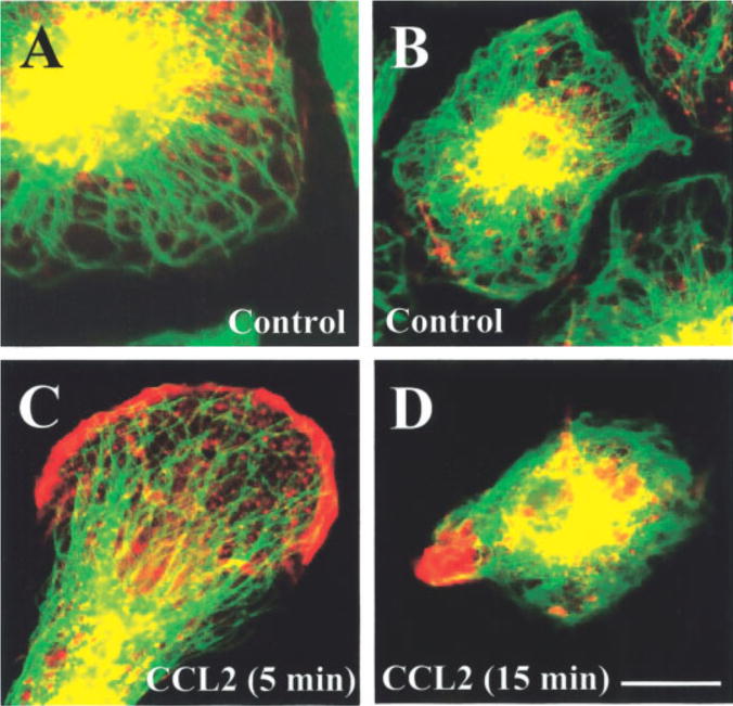
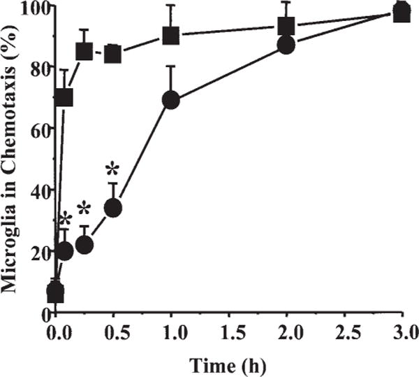
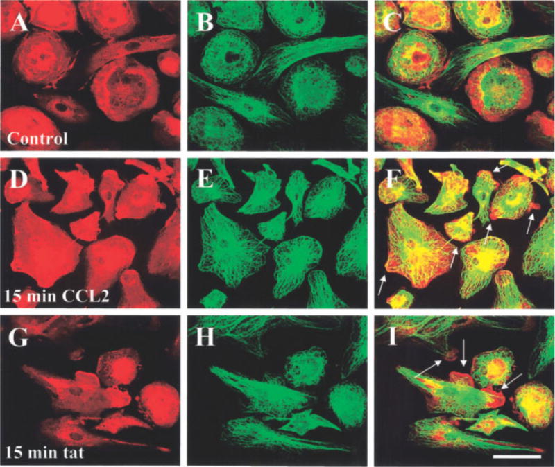
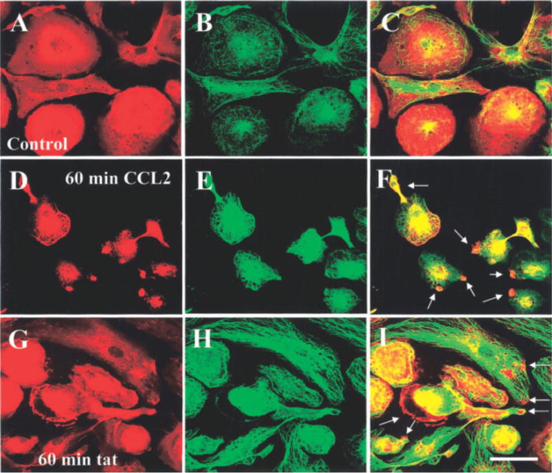
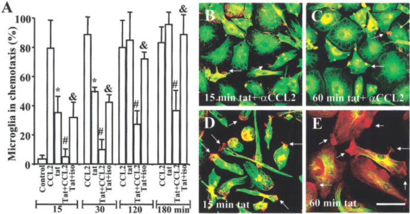
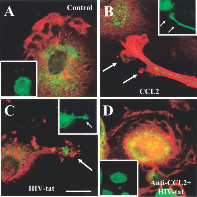
Acquired immune deficiency syndrome (AIDS) encephalitis and dementia are characterized by neuronal loss, astrogliosis, and microglia activation and migration that contribute to the formation of multinucleated giant cells. Despite extensive evidence of pathological changes in the brain of infected individuals, the mechanisms of human immune deficiency virus type 1 (HIV-1) entry, microglia migration, and viral propagation within the brain are still not completely understood. In this study, we report that the induction of a migratory phenotype in human fetal microglia by the HIV-1 transactivator protein, tat, is mediated by the chemokine, CCL2. CCL2 or tat treatment alone induced rearrangement of actin and the formation of microglial processes. The time course of cell membrane ruffling induced by CCL2 was faster (5-30 min) than that elicited by tat treatment (2-3 h). Our previous data in human fetal microglia showed that tat induces CCL2 expression. Thus, we examined whether tat-induced microglia membrane ruffling and process formation, critical components in cell migration, are mediated by the secretion of CCL2 by these cells. To test this hypothesis, we treated microglia with tat protein in the presence of neutralizing CCL2 antibodies. Co-treatment with neutralizing CCL2 antibodies resulted in the loss of tat-induced membrane ruffling. Tat treatment of microglia induced polarization of CCR2, the receptor for CCL2, to the leading edge of processes, further suggesting a CCL2-dependent mechanism of tat-induced microglia migration. Our data indicate that tat facilitates microglia migration by inducing autocrine CCL2 release. Our results suggest that tat induced CCL2 secretion may be one of the early signals during NeuroAIDS.


Figures

Fig. 1
Fig. 1
Cytoskeletal changes in human fetal microglia in response to CCL2 treatment. Microglia were stained for f-actin (Texas-red-phalloidin, red staining) and tubulin (FITC, green staining); their distribution was analyzed by confocal microscopy. A,B: Two different cultures of resting microglia (untreated), with perinuclear distribution of actin and tubulin. C: After 5 min of CCL2 treatment, early process formation and actin redistribution to the leading edge are evident. D: After 15 min of CCL2 treatment, microglia exhibit cell contraction and focal conglomeration of actin at the leading edge. Scale bar = 15 μm.
Fig. 2
Fig. 2
Time course of cytoskeletal changes and process formation in human fetal microglia after treatment with CCL2 or HIV-tat protein. The number of microglia exhibiting a migratory phenotype is presented as the percentage of microglia in chemotaxis, microglia with membrane ruffling, and process formation after different times of treatment (0, 5, 15, 30, 60, 120, and 180 min) with CCL2 (■, 100 ng/ml) or tat (●,100 ng/ml). Each value corresponds to the mean ± SD (n = 6). *P 0.005 correspond to significant differences between microglia treated with tat compared with CCL2 at the same time.
Fig. 3
Fig. 3
CCL2 or tat treatment induces rearrangement of f-actin and tubulin in human fetal microglia. Double immunofluorescence staining for f-actin (Texas Red-phalloidin, red) and tubulin (FITC staining, green) was analyzed by confocal microscopy. Representative staining for f-actin in untreated (control) cultures (A) and in cultures treated with CCL2 (100 ng/ml) (D) or tat (100 ng/ml) (G) for 15 min is shown. Representative staining for tubulin in untreated (control) cultures (B), and in cultures treated with CCL2 (100 ng/ml) (E) or tat (100 ng/ml) (H) for 15 min is also shown. Colocalization of both antigens (yellow-orange) in control (C), CCL2 (F), and tat (I)-treated microglia. Arrows denote processes induced by CCL2 or tat treatment. (n = 6) Scale bar = 40 μm.
Fig. 4
Fig. 4
CCL2 or tat treatment for 60 min induces rearrangement of f-actin and tubulin in human fetal microglia. Double immunofluorescence for f-actin (Texas Red-phalloidin, red) and tubulin (FITC staining, green) was analyzed by confocal microscopy. Representative staining is shown for f-actin in control cultures (A), and after 60 min of treatment with CCL2 (100 ng/ml) (D) or tat (100 ng/ml) (G). Tubulin staining is shown in control cultures (B), and after 60 min of treatment with CCL2 (100 ng/ml) (E) or tat (100 ng/ml) (H). Colocalization (yellow-orange) of both antigens: control (C), CCL2 (F), and tat (I)-treated microglia. Arrows denote the formation of processes induced by CCL2 or tat treatment (n = 6). Scale bar = 40 μm.
Fig. 5
Fig. 5
Neutralizing antibodies to CCL2 block the rearrangement of f-actin and tubulin induced by tat. The role of CCL2 in tat-induced cytoskeletal changes was examined using a neutralizing antibody to CCL2 (100 μg/ml). We examined the percentage of microglia exhibiting a migratory phenotype at different times points after CCL2 or tat treatment. Pre-incubation for 15 min with neutralizing CCL2 antibody (αCCL2) before tat treatment reduced the number of cells exhibiting membrane ruffling by 50–70%. The addition of a negative control, isotype-matched IgG2B myeloma protein (100 μg/ml; Iso) had no effect. Each bar corresponds to the average ±SD (n = 5). *P 0.005 corresponds to significant differences between microglia treated with CCL2 compared with tat at the same time. #P < 0.005 corresponds to significant differences between microglia treated with tat protein compared with neutralizing-CCL2 antibodies plus tat at the same time. αP 0.005 corresponds to significant differences between microglia treated with neutralizing-CCL2 antibodies plus tat compared with isotype matched antibodies plus tat at the same time. Representative pictures of f-actin (red) and tubulin (green) staining or their colocalization (orange) after 15 and 60 min of tat treatment plus neutralizing antibodies to CCL2 (B and C). Note tat-treated microglia by 15 min (D) and 60 min (E) (cf. B and C). Arrows denote the formation of processes. Scale bar = 40 μm.
Fig. 6
Fig. 6
CCL2 and tat treatment induce the redistribution of CCR2 to the edge of microglia processes. The distribution of CCR2 was determined by indirect immunofluorescence staining for f-actin (red staining) and CCR2 (green staining) in the microglia processes induced by CCL2 or tat. In control cells, CCR2 is distributed thought the cell (A), while CCL2 (B), tat (C), or anti-CCL2 plus tat (D) treatment for 15 min induces colocalization of CCR2 and actin (yellow-orange staining) in processes. The small inserts correspond to CCR2 staining alone to denote their distribution in the processes. Scale bar = 75 μm.

References

    1. Albini A, Ferrini S, Benelli R, Sforzini S, Giunciuglio D, Aluigi MG, Proudfoot AE, Alouani S, Wells TN, Mariani G, Rabin RL, Farber JM, Noonan DM. HIV-1 Tat protein mimicry of chemokines. Proc Natl Acad Sci USA. 1998;95:13153–13158. - PMC - PubMed
    1. Boddeke EW, Meigel I, Frentzel S, Gourmala NG, Harrison JK, Buttini M, Spleiss O, Gebicke-Harter P. Cultured rat microglia express functional beta-chemokine receptors. J Neuroimmunol. 1999;98:176–184. - PubMed
    1. Bretscher MS. Getting membrane flow and the cytoskeleton to cooperate in moving cells. Cell. 1996a;87:601–606. - PubMed
    1. Bretscher MS. Moving membrane up to the front of migrating cells. Cell. 1996b;85:465–467. - PubMed
    1. Brown AR, Covington M, Newton RC, Ramage R, Welch P. The total chemical synthesis of monocyte chemotactic protein-1 (MCP-1) J Pept Sci. 1996;2:40–46. - PubMed

Publication types

MeSH terms

LinkOut - more resources