. 2004 Dec 3:5:52.
doi: 10.1186/1471-2202-5-52.

Multiple synaptic and membrane sites of anesthetic action in the CA1 region of rat hippocampal slices

Affiliations

Multiple synaptic and membrane sites of anesthetic action in the CA1 region of rat hippocampal slices

Sky Pittson et al. BMC Neurosci. .

Abstract

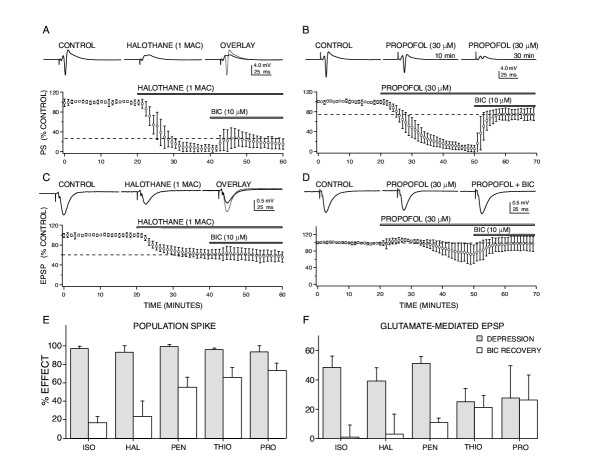
Background: Anesthesia is produced by a depression of central nervous system function, however, the sites and mechanisms of action underlying this depression remain poorly defined. The present study compared and contrasted effects produced by five general anesthetics on synaptic circuitry in the CA1 region of hippocampal slices.

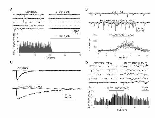
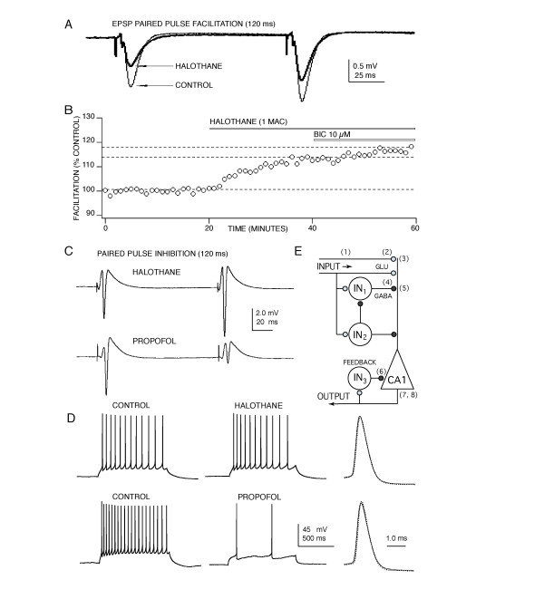
Results: At clinically relevant and equi-effective concentrations, presynaptic and postsynaptic anesthetic actions were evident at glutamate-mediated excitatory synapses and at GABA-mediated inhibitory synapses. In addition, depressant effects on membrane excitability were observed for CA1 neuron discharge in response to direct current depolarization. Combined actions at several of these sites contributed to CA1 circuit depression, but the relative degree of effect at each site was different for each anesthetic studied. For example, most of propofol's depressant effect (> 70 %) was reversed with a GABA antagonist, but only a minor portion of isoflurane's depression was reversed (< 20 %). Differences were also apparent on glutamate synapses-pentobarbital depressed transmission by > 50 %, but thiopental by only < 25 %.

Conclusions: These results, in as much as they may be relevant to anesthesia, indicate that general anesthetics act at several discrete sites, supporting a multi-site, agent specific theory for anesthetic actions. No single effect site (e.g. GABA synapses) or mechanism of action (e.g. depressed membrane excitability) could account for all of the effects produced for any anesthetic studied.


Figures

Figure 1
Figure 1
Anesthetic-induced depression of CA1 neuron responses involve actions at both glutamate and GABA-mediated synapses. (A) Halothane depressed population spike (PS) responses at clinically relevant concentrations (1 rat MAC = 1.2 vol % ~ 250 μM) and this depression was only partially reversed using a GABAA receptor antagonist, bicuculline (BIC). (B) Propofol (30 μM) produced a comparable degree of population spike depression compared to halothane, but this depression was substantially reversed with BIC, indicating that enhanced GABA-mediated inhibition contributes > 75 % of the depressant effect of propofol. The representative recordings shown are for propofol effects at 10 minutes following exposure to anesthetic (i.e. at 30 min on the x axis for the grouped data) and after 30 minutes of exposure, when a nearly complete block of the population spike was apparent. (C) Excitatory postsynaptic potentials (EPSP) were also depressed by halothane and this effect was not reversed by BIC, indicating a direct effect of the anesthetic on glutamate-mediated synapses. (D) Propofol-induced depression of glutamate-mediated EPSPs, in contrast, appeared to involve enhanced GABA-mediated inhibition, since this depression was completely reversed by BIC. The two anesthetics exhibited quite different sensitivities to reversal by BIC, indicating that actions at GABA synapses vary for these agents. For each graph, data were normalized and each point represents the mean ± SD for at least five measures from different slices made from separate animals. Horizontal bars indicate the time of exposure to each drug. Sample recordings from representative experiments are shown in the top traces. (E) BIC reversal of anesthetic-induced population spike depression was agent specific for equieffective levels of depression, and data are summarized for four anesthetics as bar graphs. Shaded bars represent the degree of depression produced by each anesthetic and open bars show the extent of reversal produced by BIC, error bars indicate SD for at least five measures from different slices. Volatile agents (isoflurane – ISO, 350 μM; halothane – HAL, 250 μM) were only weakly reversed by BIC, while intravenous agents (pentobarbital – PEN, 400 μM; thiopental – THIO, 80 μM; propofol-PRO, 30 μM) were more sensitive to the GABA receptor antagonist. (F) A similar profile of agent specific effects for BIC reversal was evident for glutamate-mediated EPSP responses, volatile agent effects were poorly reversed while intravenous agents appeared to be more sensitive to the GABA antagonist.
Figure 2
Figure 2
Whole cell patch clamp recordings of spontaneous GABA-mediated inhibitory postsynaptic currents (IPSC) in CA1 neurons revealed two sites of action for anesthetic-induced enhanced inhibition. (A) Spontaneous synaptic currents were blocked by a GABAA-receptor antagonist bicuculline (BIC), indicating that they were GABAA-mediated Cl- currents. Traces on the top show 20 s of continuous recording from a CA1 neuron in control and in the presence of BIC. A rate meter graph (bottom) shows the relatively stable frequency of IPSCs during 20 min of control recording, followed by a rapid and complete block of responses produced by BIC (indicated by the bar). (B) Halothane produced a marked increase of the inhibitory charge transfer in CA1 neurons measured as the integral of current recordings (shaded area in top traces). A three fold increase of inhibitory charge (pico Colombs – pC) was reversibly produced by halothane and appeared to come about through both a prolongation of IPSC time course (C) and from an increased frequency of synaptic currents (D). Both effects persisted in the presence of 10 μM tetrodotoxin (TTX) and/or glutamate receptor antagonists indicating that action potential dependent activity and glutamate synapses were not required for anesthetic action. For the rate meter histograms in (A) and (D), each bin represents the number of events recorded in 4 s divided by 4 to give a frequency in Hz (events/second). For all IPSC recordings a CsCl based internal solution was used in the patch pipette.
Figure 3
Figure 3
Anesthetics act at several sites to depress CA1 neuron synaptically evoked discharge. (A) Halothane appears to act presynaptically to depress glutamate release, evidenced by an increase in paired pulse facilitation concomitant with EPSP depression. A similar increase in facilitation was produced by isoflurane and pentobarbital, but not by thiopental or propofol. No change in EPSP rise or decay time was apparent in the presence any anesthetic. (B) The increased facilitation produced by halothane, isoflurane and pentobarbital was not reversed by bicuculline (BIC) indicating a depressant effect on glutamate nerve terminals – independent of anesthetic effects at GABAA receptors. (C) Differential GABA effects were also evident for paired pulse inhibition of population spike responses. Volatile agents like halothane produced little or no paired pulse inhibition at concentrations that produced a half maximal depression of first pulse responses. In contrast, propofol increased paired pulse inhibition and similar effects were observed with thiopental and pentobarbital. This increase in paired pulse inhibition was reversed by bicuculline indicating that these anesthetics enhanced recurrent GABAA-mediated inhibition. (D) The anesthetics also appeared to act directly on CA1 pyramidal neuron membrane excitability to slow action potential discharge activity, although the intravenous agents were much more effective compared to volatile anesthetics. None of the anesthetics produced an appreciable effect on individual action potential amplitude or time course (right: control – solid line; anesthetic – dotted, for halothane on top and propofol on bottom). (E) Anesthetics act at multiple sites to depress the CA1 neuron circuit. Sites of action are indicated on a diagram of CA1 circuitry showing input from Schaffer-collateral fibers, local inhibitory interneurons (IN) and a CA1 pyramidal neuron (triangle). Action potential propagation in Schaffer-collateral fibers (1) was depressed by ~ 15% by halothane and this contributes about 25 % to EPSP depression [60, 61]; see also [17]. This effect did not contribute to anesthetic-induced increases in facilitation, because no change in facilitation occurred when a comparable amount of action potential depression was produced by tetrodotoxin [61]. Further presynaptic depression at glutamate nerve terminals (2) was evident from the increased facilitation observed (Fig. 3A&3B) and there is also good evidence for postsynaptic depressant effects on both NMDA and AMPA receptors (3) [16, 19, 62, 63]. Anesthetics also act pre- and postsynaptically at GABA-mediated synapses (4, see Fig. 2) and can also increase tonic GABA-mediated inhibition by acting as GABA agonists in the absence of synaptically released GABA (5) [46-48]. Perisynaptic and extrasynaptic tonic GABAA receptors (7) also contribute to the postsynaptic depression produced by isoflurane [64] as well as thiopental and propofol [4, 65]. Enhanced recurrent inhibition (6) plays and important role for anesthetics in vivo [45] and strong effects were evident in the present study for propofol, thiopental and pentobarbital (Fig. 3C), similar to effects previously reported for halothane in hippocampal slices [26]. In addition, anesthetics also directly depress CA1 neuron excitability by blocking calcium channels and enhancing potassium currents contributing to hyperpolarization (8) and increased discharge thresholds [40, 41, 42, also Nishikawa, Beida & Maclver, unpublished]. These latter effects could influence CA1 neuron discharge activity for near threshold responses, but for the stronger stimuli used in the present study, effects on GABA and glutamate synapses and on postsynaptic receptors for these transmitters appear to contribute most (~ 80 %) to the depressant actions observed.

References

    1. Nicoll RA, Eccles JC, Oshima T, Rubia F. Prolongation of hippocampal inhibitory postsynaptic potentials by barbiturates. Nature. 1975;258:625–627. - PubMed
    1. Tanelian DL, Kosek P, Mody I, Maclver MB. The role of the GABAA receptor/chloride channel complex in anesthesia. Anesthesiology. 1993;78:757–776. - PubMed
    1. Franks NP, Lieb WR. Molecular and cellular mechanisms of general anaesthesia. Nature. 1994;367:607–614. doi: 10.1038/367607a0. - DOI - PubMed
    1. Bieda MC, Maclver MB. A Major Role For Tonic GABAA Conductances In Anesthetic Suppression Of Intrinsic Neuronal Excitability. Journal of Neurophysiology. 2004;92:1658–1667. doi: 10.1152/jn.00223.2004. - DOI - PubMed
    1. Lopes CM, Franks NP, Lieb WR. Actions of general anaesthetics and arachidonic pathway inhibitors on K+ currents activated by volatile anaesthetics and FMRFamide in molluscan neurones. British Journal of Pharmacology. 1998;125:309–318. - PMC - PubMed

Publication types

MeSH terms

LinkOut - more resources