

P80, the HinT interacting membrane protein, is a secreted antigen of Mycoplasma hominis
- PMID: 15579213
- PMCID: PMC539234
- DOI: 10.1186/1471-2180-4-46
P80, the HinT interacting membrane protein, is a secreted antigen of Mycoplasma hominis
Abstract
Background: Mycoplasmas are cell wall-less bacteria which encode a minimal set of proteins. In Mycoplasma hominis, the genes encoding the surface-localized membrane complex P60/P80 are in an operon with a gene encoding a cytoplasmic, nucleotide-binding protein with a characteristic Histidine triad motif (HinT). HinT is found in both procaryotes and eukaryotes and known to hydrolyze adenosine nucleotides in eukaryotes. Immuno-precipitation and BIACore analysis revealed an interaction between HinT and the P80 domain of the membrane complex. As the membrane anchored P80 carries an N-terminal uncleaved signal peptide we have proposed that the N-terminus extends into the cytoplasm and interacts with the cytosolic HinT.
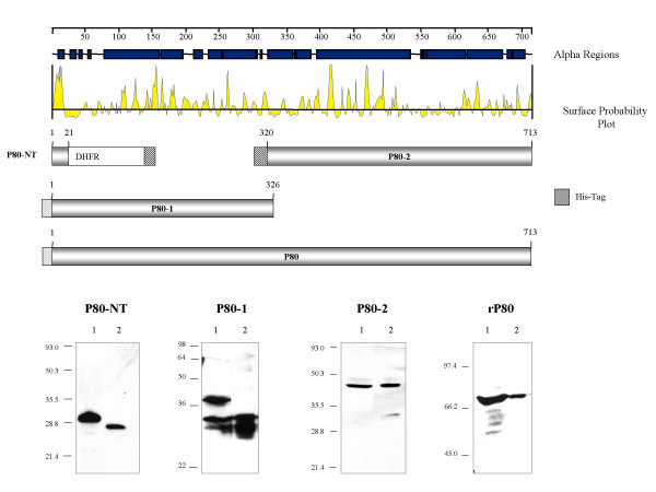
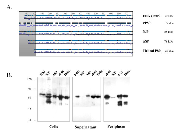
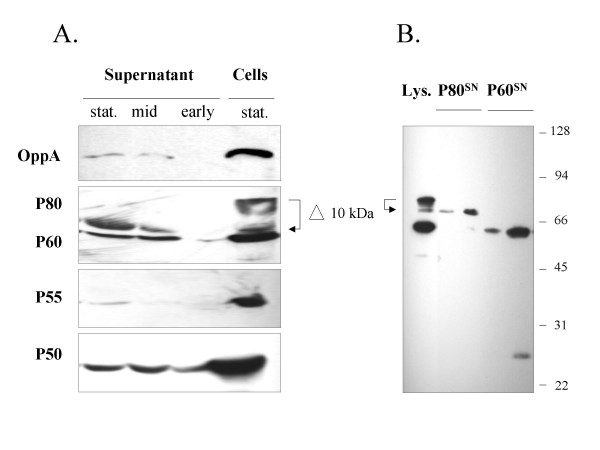
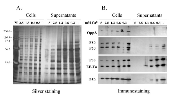
Results: Further characterization of P80 suggested that the 4.7 kDa signal peptide is protected from cleavage only in the membrane bound form. We found several proteins were released into the supernatant of a logarithmic phase mycoplasma culture, including P80, which was reduced in size by 10 kDa. Western blot analysis of recombinant P80 mutants expressed in E. coli and differing in the N-terminal region revealed that mutation of the +1 position of the mature protein (Asn to Pro) which is important for signal peptidase I recognition resulted in reduced P80 secretion. All other P80 variants were released into the supernatant, in general as a 74 kDa protein encompassing the helical part of P80. Incubation of M. hominis cells in phosphate buffered saline supplemented with divalent cations revealed that the release of mycoplasma proteins into the supernatant was inhibited by high concentrations of calciumions.
Conclusions: Our model for secretion of the P80 protein of M. hominis implies a two-step process. In general the P80 protein is transported across the membrane and remains complexed to P60, surface-exposed and membrane anchored via the uncleaved signal sequence. Loss of the 4.7 kDa signal peptide seems to be a pre-requisite for P80 secretion, which is followed by a proteolytic process leading to a helical 74 kDa product. We propose that this novel form of two-step secretion is one of the solutions to a life with a reduced gene set.
Figures
Similar articles
-
The cytosolic HinT protein of Mycoplasma hominis interacts with two membrane proteins.Mol Microbiol. 2001 Jul;41(1):279-87. doi: 10.1046/j.1365-2958.2001.02524.x. Mol Microbiol. 2001. PMID: 11454219
-
HinT proteins and their putative interaction partners in Mollicutes and Chlamydiaceae.BMC Microbiol. 2005 May 18;5:27. doi: 10.1186/1471-2180-5-27. BMC Microbiol. 2005. PMID: 15904496 Free PMC article.
-
Production of a chimeric protein and its potential application in sero-diagnosis of Mycoplasma hominis infection.J Microbiol Methods. 2018 Jan;144:186-191. doi: 10.1016/j.mimet.2017.12.001. Epub 2017 Dec 5. J Microbiol Methods. 2018. PMID: 29217154
-
Molecular dissection of Mycoplasma hominis.APMIS Suppl. 2000;97:1-45. APMIS Suppl. 2000. PMID: 10721331 Review.
-
Molecular biology of Mycoplasma.Wien Klin Wochenschr. 1997 Aug 8;109(14-15):557-61. Wien Klin Wochenschr. 1997. PMID: 9286059 Review.
Cited by
-
Paradigms of Protist/Bacteria Symbioses Affecting Human Health: Acanthamoeba species and Trichomonas vaginalis.Front Microbiol. 2021 Jan 7;11:616213. doi: 10.3389/fmicb.2020.616213. eCollection 2020. Front Microbiol. 2021. PMID: 33488560 Free PMC article. Review.
-
Being pathogenic, plastic, and sexual while living with a nearly minimal bacterial genome.PLoS Genet. 2007 May 18;3(5):e75. doi: 10.1371/journal.pgen.0030075. PLoS Genet. 2007. PMID: 17511520 Free PMC article.
-
Novel Secreted Protein of Mycoplasma bovis MbovP280 Induces Macrophage Apoptosis Through CRYAB.Front Immunol. 2021 Feb 15;12:619362. doi: 10.3389/fimmu.2021.619362. eCollection 2021. Front Immunol. 2021. PMID: 33659004 Free PMC article.
-
Mycoplasma bovis Membrane Protein MilA Is a Multifunctional Lipase with Novel Lipid and Glycosaminoglycan Binding Activity.Infect Immun. 2020 May 20;88(6):e00945-19. doi: 10.1128/IAI.00945-19. Print 2020 May 20. Infect Immun. 2020. PMID: 32253247 Free PMC article.
-
Survey of surface proteins from the pathogenic Mycoplasma hyopneumoniae strain 7448 using a biotin cell surface labeling approach.PLoS One. 2014 Nov 11;9(11):e112596. doi: 10.1371/journal.pone.0112596. eCollection 2014. PLoS One. 2014. PMID: 25386928 Free PMC article.
References
Publication types
MeSH terms
Substances
LinkOut - more resources
Full Text Sources
Other Literature Sources
Research Materials
