. 2004 Jul-Aug;21(4):545-50.
doi: 10.1017/S0952523804214055.

N-type and L-type calcium channels mediate glycinergic synaptic inputs to retinal ganglion cells of tiger salamanders

Affiliations

N-type and L-type calcium channels mediate glycinergic synaptic inputs to retinal ganglion cells of tiger salamanders

Mark C Bieda et al. Vis Neurosci. 2004 Jul-Aug.

Abstract

Synaptically localized calcium channels shape the timecourse of synaptic release, are a prominent site for neuromodulation, and have been implicated in genetic disease. In retina, it is well established that L-type calcium channels play a major role in mediating release of glutamate from the photoreceptors and bipolar cells. However, little is known about which calcium channels are coupled to synaptic exocytosis of glycine, which is primarily released by amacrine cells. A recent report indicates that glycine release from spiking AII amacrine cells relies exclusively upon L-type calcium channels. To identify calcium channel types controlling neurotransmitter release from the population of glycinergic neurons that drive retinal ganglion cells, we recorded electrical and potassium evoked inhibitory synaptic currents (IPSCs) from these postsynaptic neurons in retinal slices from tiger salamanders. The L-channel antagonist nifedipine strongly inhibited release and FPL64176, an L-channel agonist, greatly enhanced it, indicating a significant role for L-channels. omega-Conotoxin MVIIC, an N/P/Q-channel antagonist, strongly inhibited release, indicating an important role for non-L channels. While the P/Q-channel blocker omega-Aga IVA produced only small effects, the N-channel blocker omega-conotoxin GVIA strongly inhibited release. Hence, N-type and L-type calcium channels appear to play major roles, overall, in mediating synaptic release of glycine onto retinal ganglion cells.


Figures
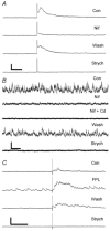
Fig. 1
Fig. 1
L-channels mediate release of glycine. A: Nifedipine (20 μM) strongly suppresses the electrically evoked IPSC (eIPSC) in a mostly reversible manner. Traces are mean of three consecutive responses per condition. B: Nifedipine (50 μM) suppresses release induced by continuous application of 20 mM K+-saline (HK-sIPSCs). Cadmium (100 μM) suppresses almost all nifedipine-insensitive release. Raw sweeps (6 s) are shown. C: FPL64176 (2 μM), a L-channel agonist, enhances the eIPSC and baseline transmission in a partially reversible manner. Traces are mean of four consecutive responses per condition. Scale bars: A = 30 pA, 300 ms; B = 20 pA, 500 ms; and C = 30 pA, 500 ms. Con: control; Nif: nifedipine; Nif + Cd: Nifedipine + cadmium; Wash: post-drug washout; and Strych: strychnine (10 μM); and FPL: FPL64176.
Fig. 2
Fig. 2
ω-conotoxin MVIIC (5 μM), an N/P/Q antagonist, suppresses release of glycine. A: MVIIC suppresses the majority of glycine release. Traces are mean of 3/condition. B: MVIIC suppresses release induced by continuous application of 20 mM K+-saline. MVIIC-resistant release is cadmium (100 μM) sensitive. Traces are raw 60-s sweeps. Scale bars: A = 30 pA, 500 ms; and B = 25 pA, 10 s. MVIIC: ω-conotoxin MVIIC (5 μM); others are as in Fig. 1.
Fig. 3
Fig. 3
P/Q channels play a small role in mediating glycine release. A: ω-Aga IVa (200 nM) has little effect on eIPSC. Traces are mean of 3/condition. B: ω-Aga IVa (200 nM) has small effects on release induced by continuous application of 20 mM K+-saline. Cadmium (75 μM) mostly reversibly blocks release. Traces are raw 60-s sweeps. Scale bars: A = 40 pA, 800 ms; and B = 15 pA, 10 s. Aga: ω-Aga IVa; others are as in Fig. 1.
Fig. 4
Fig. 4
N-channels play a major role in release of glycine. A: ω-conotoxin GVIA (1 μM) strongly suppresses eIPSC. Traces shown have strychnine-insensitive electrical artifact digitally subtracted for clarity (see Results). Traces are mean of 3/condition. B: GVIA (2 μM) strongly suppresses release induced by continuous application of 20 mM K+-saline. Cell was voltage clamped at -60 mV with high chloride internal solution. Events are inward (downward) currents. In this recording, cadmium (100 μM) and strychnine (10 μM) induce little additional block. Traces are raw 60-s sweeps. Scale bars: A = 20 pA, 800 ms; and B = 30 pA, 10 s. GVIA: ω-conotoxin GVIA; others are as in Fig. 1.

References

    1. Barnes S, Werblin F. Gated currents generate single spike activity in amacrine cells of the tiger salamander retina. Proceedings of the National Academy of Sciences of the U.S.A. 1986;83:1509–1512. - PMC - PubMed
    1. Bieda MC, Copenhagen DR. Sodium action potentials are not required for light-evoked release of GABA or glycine from retinal amacrine cells. Journal of Neurophysiology. 1999;81:3092–3095. - PubMed
    1. Bieda MC, Copenhagen DR. Inhibition is not required for the production of transient spiking responses from retinal ganglion cells. Visual Neuroscience. 2000;17:243–254. - PubMed
    1. Burgess DL, Noebels JL. Voltage-dependent calcium channel mutations in neurological disease. Annals of the New York Academy of Sciences. 1999;868:199–212. - PubMed
    1. Cook PB, Lukasiewicz PD, McReynolds JS. Action potentials are required for the lateral transmission of glycinergic transient inhibition in the amphibian retina. Journal of Neuroscience. 1998;18:2301–2308. - PMC - PubMed

Publication types

MeSH terms