. 2004 Nov;23(7):421-32.
doi: 10.1016/j.matbio.2004.09.007.

Homozygous ablation of fibroblast growth factor-23 results in hyperphosphatemia and impaired skeletogenesis, and reverses hypophosphatemia in Phex-deficient mice

Affiliations

Homozygous ablation of fibroblast growth factor-23 results in hyperphosphatemia and impaired skeletogenesis, and reverses hypophosphatemia in Phex-deficient mice

Despina Sitara et al. Matrix Biol. 2004 Nov.

Abstract

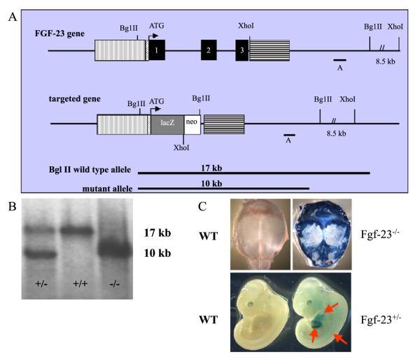
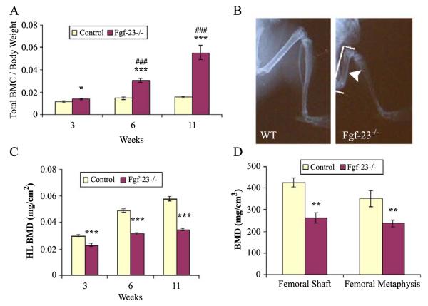
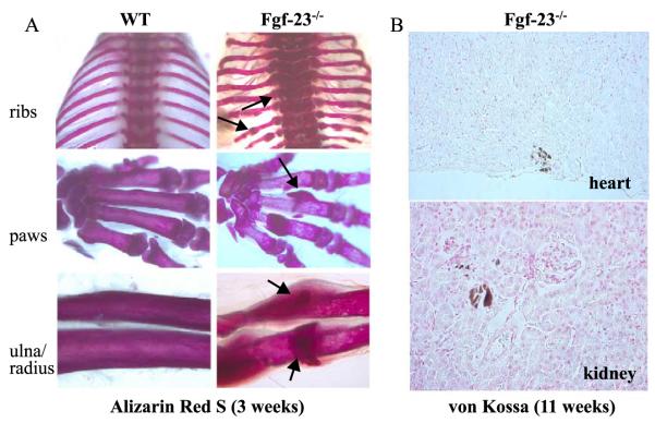
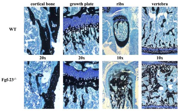
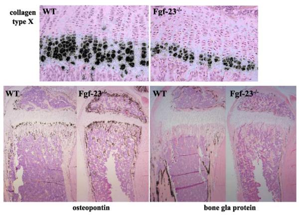
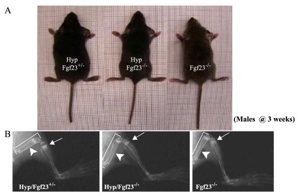
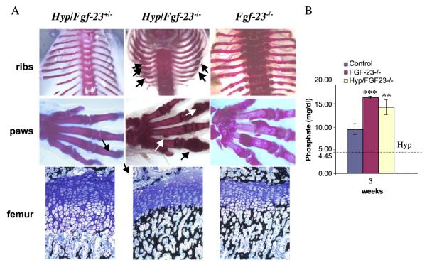
Fibroblast growth factor-23 (FGF-23), a recently identified molecule that is mutated in patients with autosomal dominant hypophosphatemic rickets (ADHR), appears to be involved in the regulation of phosphate homeostasis. Although increased levels of circulating FGF-23 were detected in patients with different phosphate-wasting disorders such as oncogenic osteomalacia (OOM) and X-linked hypophosphatemia (XLH), it is not yet clear whether FGF-23 is directly responsible for the abnormal regulation of mineral ion homeostasis and consequently bone development. To address some of these unresolved questions, we generated a mouse model, in which the entire Fgf-23 gene was replaced with the lacZ gene. Fgf-23 null (Fgf-23-/-) mice showed signs of growth retardation by day 17, developed severe hyperphosphatemia with elevated serum 1,25(OH)2D3 levels, and died by 13 weeks of age. Hyperphosphatemia in Fgf-23-/- mice was accompanied by skeletal abnormalities, as demonstrated by histological, molecular, and various other morphometric analyses. Fgf-23-/-) mice had increased total-body bone mineral content (BMC) but decreased bone mineral density (BMD) of the limbs. Overall, Fgf-23-/- mice exhibited increased mineralization, but also accumulation of unmineralized osteoid leading to marked limb deformities. Moreover, Fgf-23-/- mice showed excessive mineralization in soft tissues, including heart and kidney. To further expand our understanding regarding the role of Fgf-23 in phosphate homeostasis and skeletal mineralization, we crossed Fgf-23-/- animals with Hyp mice, the murine equivalent of XLH. Interestingly, Hyp males lacking both Fgf-23 alleles were indistinguishable from Fgf-23/-/ mice, both in terms of serum phosphate levels and skeletal changes, suggesting that Fgf-23 is upstream of the phosphate regulating gene with homologies to endopeptidases on the X chromosome (Phex) and that the increased plasma Fgf-23 levels in Hyp mice (and in XLH patients) may be at least partially responsible for the phosphate imbalance in this disorder.


Figures

Fig. 1
Fig. 1
(A) Schematic representation of the murine Fgf-23 gene and the corresponding knock-out/in targeting vector. Exons 1 to 3 are shown in black boxes. Vertical and horizontal shaded boxes represent the 5′ and 3′ flanking regions of the Fgf-23 gene, respectively, which were used for homologous recombination. The lacZ gene was cloned in frame with the initiator methionine of the Fgf-23 gene. The neomycin resistance (neo) gene is driven by the phosphoglycerate kinase-1 (PGK-1) promoter and contains an Sv40 polyA adenylation site. Probe A was used as external probe to hybridize genomic Southern blots (BglII digest) shown in (B) (wild type=+/+, heterozygous=+/−, homozygous=−/−). Panel (C) represents lacZ staining of a wild-type (lower left) and a heterozygous Fgf-23 embryo (Fgf-23+/−, lower right) at E12.5. Arrows depict lacZ positive tissues (somites, liver, and heart). Upper panels demonstrate lacZ staining in a wild type (left) and Fgf-23−/− (right) skull at 3 weeks. Blue staining represents expression of the Fgf-23 gene.
Fig. 2
Fig. 2
(A) Graphic display of total bone mineral content (BMC) of control and Fgf-23−/− animals at 3, 6, and 11 weeks. Each value obtained for BMC was normalized to the body weight of the corresponding animal. Fgf-23−/− mice show a statistical significant increase in total BMC when compared to control littermates (*=p<0.05; ***=p<0.0001). A statistically significant increase in total BMC was also observed among Fgf-23−/− mice with time (###=p<0.0001). (B) X-ray autoradiography of hindlimbs from a wild-type (WT) and an Fgf-23−/− mouse. Brackets depict length, and arrowhead depicts thickness of femur in Fgf-23−/− mouse. (C) Graph represents bone mineral density of hindlimbs measured by PIXImus analysis. Fgf-23−/− mice show a statistically significant decrease in BMD at 3, 6, and 11 weeks when compared to controls (***=p<0.0001). (D) BMD obtained from femoral shaft (left) and femoral methaphysis (right) of wild-type (white bar) and Fgf-23−/− animals (dark bar) by QCT at 4 weeks of age. Fgf-23−/− mice show a statistically significant decrease in BMD (**=p<0.001).
Fig. 3
Fig. 3
(A) Alizarin red S staining of skeletal elements (ribs, paws, ulna/radius) from a wild type (left panels) and Fgf-23−/− (right panels) at 3 weeks. Arrows depict some areas with abnormal mineralization in Fgf-23−/− bones. (B) Abnormal mineralization is shown in heart (top) and in and around the tubules of Fgf-23−/− kidney (bottom) at 11 weeks. (For interpretation of the references to colour in this figure legend, the reader is referred to the web version of this article.)
Fig. 4
Fig. 4
Three-micrometer-thick undecalcified sections from 4-week-old wild-type (upper panels) and Fgf-23−/− (lower panels) bones (cortical bone, growth plate, ribs, vertebra) were stained with von Kossa/McNeal (magnification 20×, 10×). Black staining represents mineralization. More mineral deposition is found in the area below the growth plate (methaphysis), ribs, and in vertebra. In contrast, areas of unmineralized osteoid (light blue) are found in cortical bone. (For interpretation of the references to colour in this figure legend, the reader is referred to the web version of this article.)
Fig. 5
Fig. 5
In situ hybridization was performed on 6μm-thick decalcified paraffin sections from tibia of wild-type (WT) and Fgf-23−/− animals at 3 weeks. The zone of hypertrophic chondrocytes was reduced in Fgf-23−/−, which was confirmed by decreased collagen type X mRNA expression. In contrast, osteopontin mRNA expression was elevated in osteoblasts of Fgf-23−/− animals, while bone gla protein (osteocalcin) mRNA expression was diminished.
Fig. 6
Fig. 6
(A) Gross features of three male littermates at 3 weeks. Shown are Hyp/Fgf23+/− (left), Fgf23−/− (right), and compound mutants Hyp/Fgf23−/− (middle). (B) X-ray autoradiographs of hindlimbs from the same animals shown in panel (A). Arrows point to the growth plate of tibia. The features typical of rickets shown in Hyp/Fgf23+/− (left panel) had improved considerably in compound mutants Hyp/Fgf23−/− (middle panel). Arrowheads depict thickness of femoral shaft of these animals. Hyp/Fgf23−/− compound mutants (middle) exhibit longer (brackets) and thinner long bones than Hyp/Fgf23+/− (left) animals.
Fig. 7
Fig. 7
(A) Alizarin red S staining of skeletal elements (ribs, paws) from a Hyp/Fgf23+/− (left), Hyp/Fgf23−/− (middle), and Fgf23−/− (right) mice at 3 weeks. Arrows depict some areas with abnormal mineralization in Hyp/Fgf23−/− bones resembling the phenotype of Fgf23−/− skeleton. Lower panels represent 3μm-thick undecalcified sections from femur of 3-week-old Hyp/Fgf23+/− (left), Hyp/Fgf23−/− (middle), and Fgf23−/− (right) mice stained with Von Kossa/McNeal. Panel (B) represents a graph comparing serum phosphate levels of wild-type controls, Fgf23−/−, and Hyp/Fgf23−/− compound mutants at 3 weeks of age. The horizontal dotted line illustrates published serum phosphate levels in Hyp mice (mean: 4.45 mg/dl) (***=p<0.0001; **=p<0.001) (Lorenz-Depiereux et al., 2004).

References

    1. ADHR_Consortium Autosomal dominant hypophosphataemic rickets is associated with mutations in FGF23. The ADHR Consortium. Nat. Genet. 2000;26:345–348. - PubMed
    1. Alfrey AC, LeGendre GR, Kaehny WD. The dialysis encephalopathy syndrome. Possible aluminum intoxication. N. Engl. J. Med. 1976;294:184–188. - PubMed
    1. Aono Y, Shimada T, Yamaziki Y, Hino R, Takeuchi Y, Fujita T, Fukumoto S, Nagano N, Wada M, Yamashita T. The neutralization of FGF-23 ameliorates hypophosphatemia and rickets in Hyp mice. J. Bone Miner. Res. 2003;18(Suppl. 2):1056.
    1. Argiro L, Desbarats M, Glorieux FH, Ecarot B. Mepe, the gene encoding a tumor-secreted protein in oncogenic hypophosphatemic osteomalacia, is expressed in bone. Genomics. 2001;74:342–351. - PubMed
    1. Bai X, Miao D, Panda D, Grady S, McKee MD, Goltzman D, Karaplis AC. Partial rescue of the Hyp phenotype by osteoblast-targeted PHEX (phosphate-regulating gene with homologies to endopeptidases on the X chromosome) expression. Mol. Endocrinol. 2002;16:2913–2925. - PubMed

Publication types

MeSH terms