Cell tropism of simian immunodeficiency virus in culture is not predictive of in vivo tropism or pathogenesis
- PMID: 15579453
- PMCID: PMC1618703
- DOI: 10.1016/S0002-9440(10)63261-0
Cell tropism of simian immunodeficiency virus in culture is not predictive of in vivo tropism or pathogenesis
Abstract
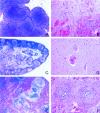
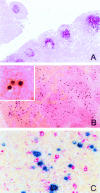
SIVmac239/316 is a molecular clone derived from SIVmac239 that differs from the parental virus by nine amino acids in env. This virus, unlike the parental SIVmac239, is able to replicate well in alveolar macrophages in culture. We have not however, observed macrophage-associated inflammatory disease in any animal infected with SIVmac239/316. Therefore, we sought to examine the cell tropism of this virus in vivo in multiple tissues using in situ hybridization combined with immunohistochemistry and multilabel confocal microscopy for viral nucleic acid and multiple cell-type-specific markers for macrophages and T lymphocytes. Tissues examined included brain, heart, lung, lymph nodes, spleen, thymus, and small and large intestine. Matched tissues from macaques infected with the parental SIVmac239 and uninfected macaques were also examined. Many infected cells were detected in the tissues of animals infected with SIVmac239 and SIVmac239/316 although there appeared to be fewer positive cells in animals infected with SIVmac239/316. Surprisingly, in light of the cell culture observations, nearly every simian immunodeficiency virus-infected cell in animals inoculated with SIVmac239/316 was a T lymphocyte rather than a macrophage. This was true both during early infection (first 2 months) and in terminal disease. In contrast, as previously described, SIVmac239 was found in both T cells and macrophages in tissues as early as 21 days after infection. These studies indicate that during both acute and chronic SIVmac239/316 infection T lymphocytes rather than macrophages are the principal targets in vivo. These data combined with the absence of macrophage-associated lesions in SIVmac239/316-infected animals indicate that in vitro cell tropism is not predictive of in vivo tropism or disease pathogenesis.
Figures


References
-
- Sasseville VG, Desrosiers RC, Morrison HG. Simian immunodeficiency viruses (retroviridae). Webster R, Granoff A, editors. London: Academic Press; 1999:pp 1640–1647.
-
- Daniel MD, Letvin NL, King NW, Kannagi M, Sehgal PK, Hunt RD, Kanki PJ, Essex M, Desrosiers RC. Isolation of T-cell tropic HTLV-III-like retrovirus from macaques. Science. 1985;228:1201–1204. - PubMed
-
- Chalifoux LV, King NW, Daniel MD, Kannagi M, Desrosiers RC, Sehgal PK, Waldron LM, Hunt RD, Letvin NL. Lymphoproliferative syndrome in an immunodeficient rhesus monkey naturally infected with an HTLV-III-like virus (STLV-III). Lab Invest. 1986;55:43–50. - PubMed
-
- Chalifoux LV, King NW, Letvin NL. Morphologic changes in lymph nodes of macaques with an immunodeficiency syndrome. Lab Invest. 1984;51:22–26. - PubMed
Publication types
MeSH terms
Substances
Grants and funding
LinkOut - more resources
Full Text Sources
Other Literature Sources

