Comparative Study
. 2004 Dec 1;561(Pt 2):547-57.
doi: 10.1113/jphysiol.2004.075168. Epub 2004 Oct 7.

Heteromeric KCNE2/KCNQ1 potassium channels in the luminal membrane of gastric parietal cells

Affiliations
Comparative Study

Heteromeric KCNE2/KCNQ1 potassium channels in the luminal membrane of gastric parietal cells

Dirk Heitzmann et al. J Physiol. .

Abstract

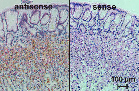
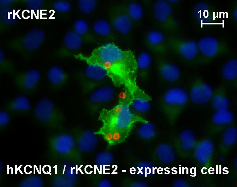
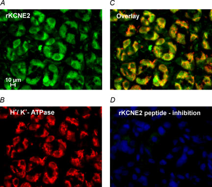
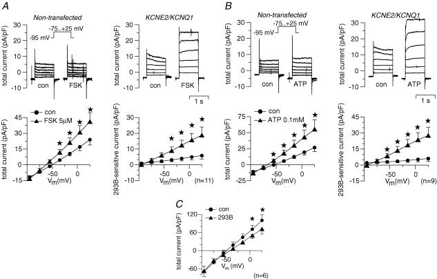
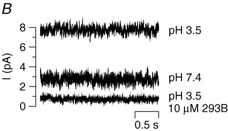
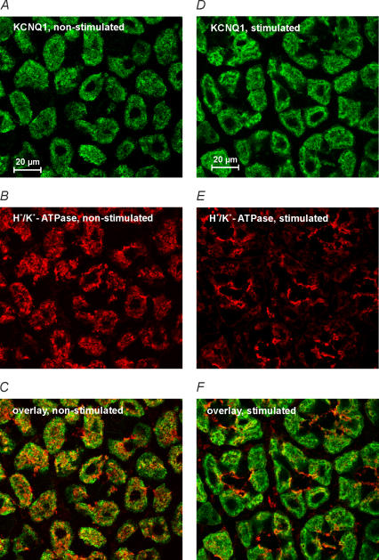
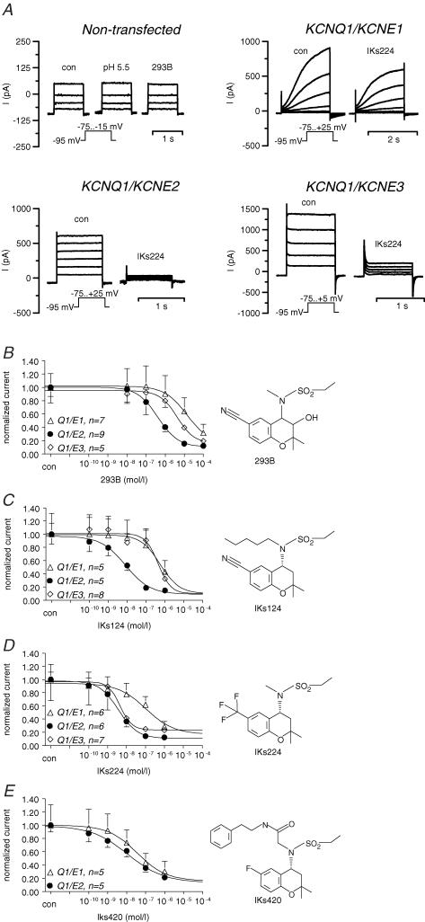
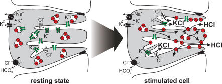
Recently, we and others have shown that luminal K+ recycling via KCNQ1 K+ channels is required for gastric H+ secretion. Inhibition of KCNQ1 by the chromanol 293B strongly diminished H+ secretion. The present study aims at clarifying KCNQ1 subunit composition, subcellular localization, regulation and pharmacology in parietal cells. Using in situ hybridization and immunofluorescence techniques, we identified KCNE2 as the beta subunit of KCNQ1 in the luminal membrane compartment of parietal cells. Expressed in COS cells, hKCNE2/hKCNQ1 channels were activated by acidic pH, PIP2, cAMP and purinergic receptor stimulation. Qualitatively similar results were obtained in mouse parietal cells. Confocal microscopy revealed stimulation-induced translocation of H+,K+-ATPase from tubulovesicles towards the luminal pole of parietal cells, whereas distribution of KCNQ1 K+ channels did not change to the same extent. In COS cells the 293B-related substance IKs124 blocked hKCNE2/hKCNQ1 with an IC50 of 8 nM. Inhibition of hKCNE1- and hKCNE3-containing channels was weaker with IC50 values of 370 and 440 nM, respectively. In conclusion, KCNQ1 coassembles with KCNE2 to form acid-activated luminal K+ channels of parietal cells. KCNQ1/KCNE2 is activated during acid secretion via several pathways but probably not by targeting of the channel to the membrane. IKs124 could serve as a leading compound in the development of subunit-specific KCNE2/KCNQ1 blockers to treat peptic ulcers.


Figures

Figure 1
Figure 1. KNCE2 in situ hybridization on human gastric mucosa
Silver grain labelling of parietal cells was detected in antisense probe-treated sections but not in sense controls.
Figure 2
Figure 2. Antibody labelling of COS cells co-transfected with rat KCNE2 and human KCNQ1
KCNE2-specific staining is shown in the central area (polyclonal, affinity-purified antibody); within this area anti-CD8 beads (circles) indicate transfected cells. HOE33342 nuclear staining is in the background.
Figure 3
Figure 3. KCNE2 in non-stimulated rat gastric mucosa
A, epifluorescence images of rat oxyntic mucosa stained with polyclonal, affinity-purified KCNE2 antibody. B, H+,K+-ATPase labelling with monoclonal antibody. C, overlay of KCNE2 and H+,K+-ATPase staining shows partial co-localization in parietal cells as described for KCNQ1 and H+,K+-ATPase. D, no staining was observed with the KCNE2 antibody after absorption to the peptide used for immunization; nuclear staining with HOE33342 is visible.
Figure 4
Figure 4. Whole-cell patch-clamp measurements of hKCNE2/hKCNQ1-transfected COS cells (A and B) and mouse parietal cells (C)
Cells were clamped stepwise from –95 to +25 mV. A, effect of forskolin (FSK) on a non-transfected cell (whole-cell current normalized to cell capacitance; left upper panel) and a KCNE2/KCNQ1 co-transfected cell (right upper panel). Left lower panel shows the summary of the FSK effect on whole-cell current in KCNE2/KCNQ1 co-transfected cells. Note the shift of reversal potential of the whole-cell current indicating FSK-induced hyperpolarization of the cells. Right lower panel shows the effect of FSK on the 293B-sensitive current as a measure of KCNE2/KCNQ1-specific current. B, effect of ATP on whole-cell current and 293B-sensitve current. Arrangement of panels as described for A. C, effect of 293B on whole-cell current of freshly isolated mouse parietal cells in the continuous presence of forskolin (5 μm) plus carbachol (100 μm). Error bars: s.e.m. Stars: significantly different from control (con). Number of experiments shown in parentheses.
Figure 5
Figure 5. Effect of acidic pH on cytosolic [Ca2+] and KCNQ1/KCNE2 induced current
A, effect of acidic extracellular pH and ionomycin (0.1 μm) on intracellular Ca2+ activity of COS cells as measured by fura-2 fluorescence (n = 10). For comparison, the peak value of ATP (100 μm, pH 7.4) is depicted. Error bars: s.e.m. Stars: significantly different from control. B, original current recordings of an outside-out patch of COS cells co-transfected with hKCNE2/hKCNQ1 (Vc = 0 mV, pipette and bath solution as described in Methods). Note the increase in current due to acidic pH and the almost complete inhibition by 293B.
Figure 6
Figure 6. Effect of parietal cell activation on the subcellular localization of KCNQ1 and H+, K+-ATPase
Confocal images of mouse gastric mucosa: A–C non-stimulated; D–F after stimulation with pentagastrin plus histamine plus carbachol. A–C: KCNQ1 (A) partially co-localized with H+,K+-ATPase (B) in non-stimulated tissue. Overlay is shown in C. D–F: activation of acid secretion did slightly change the distribution of KCNQ1 (D) but led to a marked targeting of H+,K+-ATPase to the luminal pole of parietal cells (E). Overlay is shown in F.
Figure 7
Figure 7. Pharmacology of hKCNE1/hKCNQ1 (Q1/E1), hKCNE2/hKCNQ1 (Q1/E2) and hKCNE3/hKCNQ1 (Q1/E3) expressed in COS cells
A, typical biophysical and pharmacological properties of the ‘cardiac’ KCNE1/KCNQ1 channel complex heteromeric KCNE2/KCNQ1 and KCNE3/KCNQ1 channels (Dedek & Waldegger, 2001). Currents of non-transfected cells are shown as control. Note the slow activation kinetics of KCNE1/KCNQ1 and the linear current–voltage relationship of KCNE2/KCNQ1 and KCNE3/KCNQ1. IKs224 (0.1 μm) was more effective in blocking KCNE2/KCNQ1 compared to KCNE1/KCNQ1 and KCNE3/KCNQ1 currents. For concentration–response curves, 293B (B), IKs124 (C), IKs224 (D) and IKs420 (E) were applied in rising concentrations to the bath solution. For calculations, whole-cell currents (I) at the most depolarized voltage step were normalized to control values. Error bars: s.e.m.; number of experiments are indicated in the panels.
Figure 8
Figure 8. Putative model of H+, K+-ATPase and K+ channel activation during gastric acid secretion
In the resting state of the parietal cell, H+,K+-ATPase is located in so-called tubulovesicles beneath the luminal membrane and is functionally silent. At the present time, it is not known whether H+,K+-ATPase and K+ channels are located in the same tubulovesicles. Upon stimulation of gastric acid secretion, K+ channels (probably KCNE2/KCNQ1, KCNJ1, KCNJ2 and KCNJ10) are activated via cAMP and other pathways, resulting in KCl secretion. H+,K+-ATPase is predominantly targeted to the luminal pole of canaliculi, where it pumps out H+ in exchange for K+.

References

    1. Abbott GW, Sesti F, Splawski I, Buck ME, Lehmann MH, Timothy KW, Keating MT, Goldstein SA. MiRP1 forms IKr potassium channels with HERG and is associated with cardiac arrhythmia. Cell. 1999;97:175–187. - PubMed
    1. Aihara T, Nakamura E, Amagase K, Tomita K, Fujishita T, Furutani K, Okabe S. Pharmacol Ther. 2003;98:109–127. - PubMed
    1. Angelo K, Jespersen T, Grunnet M, Nielsen MS, Klaerke DA, Olesen SP. KCNE5 induces time- and voltage-dependent modulation of the KCNQ1 current. Biophys J. 2002;83:1997–2006. - PMC - PubMed
    1. Barhanin J, Lesage F, Guillemare E, Fink M, Lazdunski M, Romey G. KvLQT1 and IsK (minK) proteins associate to form the IKs cardiac potassium current. Nature. 1996;384:78–80. - PubMed
    1. Chow DC, Forte JG. Characterization of the beta-subunit of the H+-K+-ATPase using an inhibitory monoclonal antibody. Am J Physiol. 1993;265:C1562–C1570. - PubMed

Publication types

MeSH terms

LinkOut - more resources