. 2005 Jan;137(1):127-40.
doi: 10.1104/pp.104.049361. Epub 2004 Dec 3.

The role of the cell cycle machinery in resumption of postembryonic development

Affiliations

The role of the cell cycle machinery in resumption of postembryonic development

Rosa Maria Barrôco et al. Plant Physiol. 2005 Jan.

Abstract

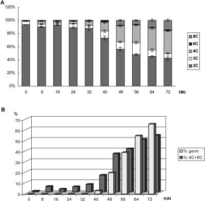
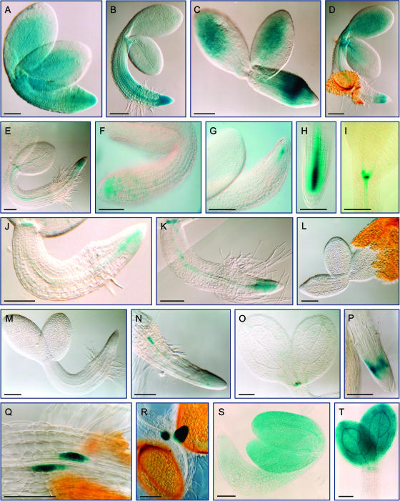
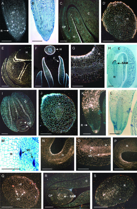
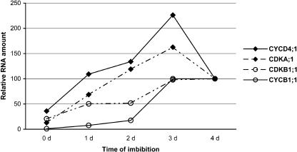
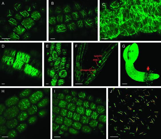
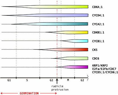
Cell cycle activity is required for plant growth and development, but its involvement in the early events that initiate seedling development remains to be clarified. We performed experiments aimed at understanding when cell cycle progression is activated during seed germination, and what its contribution is for proper seedling establishment. To this end, the spatial and temporal expression profiles of a large set of cell cycle control genes in germinating seeds of Arabidopsis (Arabidopsis thaliana) and white cabbage (Brassica oleracea) were analyzed. The in vivo behavior of the microtubular cytoskeleton was monitored during Arabidopsis seed germination. Flow cytometry of Arabidopsis germinating seeds indicated that DNA replication was mainly initiated at the onset of root protrusion, when germination reached its end. Expression analysis of cell cycle genes with mRNA in situ localization, beta-glucuronidase assays, and semiquantitative reverse transcription-polymerase chain reaction showed that transcription of most cell cycle genes was detected only after completion of germination. In vivo green fluorescent protein analysis of the microtubule cytoskeleton demonstrated that mitosis-specific microtubule arrays occurred only when the radicle had started to protrude, although the assembly of the microtubular cytoskeleton was promptly activated once germination was initiated. Thus, seed germination involves the synthesis and/or activation of a reduced number of core cell cycle proteins, which only trigger DNA replication, but is not sufficient to drive cells into mitosis. Mitotic divisions are observed only after the radicle has protruded and presumably rely on the de novo production of other cell cycle regulators.


Figures

Figure 1.
Figure 1.
A, Percentage of cells from germinating Arabidopsis seeds displaying a 2C, 3C, 4C, 6C, or 8C nuclear DNA content. B, Percentage of embryo cells from which the DNA has gone through replication [calculated as the ratio (4C + 8C)/(2C + 4C + 8C)]. In parallel, the percentage of seeds whose radicle had protruded at the defined time points is presented.
Figure 2.
Figure 2.
Promoter activity of cell cycle genes in Arabidopsis seeds and young seedlings as revealed by GUS enzymatic assays. A and B, Promoter activity of CDKA;1 in seeds imbibed for 1 and 2 d, respectively. C to E, Histochemical GUS detection of CYCA2;1 promoter activity in seeds imbibed for 1, 2, and 3 d, respectively. F to I, GUS expression driven by the CYCD4;1 promoter in dry seeds (F), and seeds imbibed for 2 (G) or 3 d (H and I). J and K, CDKB1;1 promoter activity in young seedlings (2 DAI and 3 DAI, respectively). L to R, CYCB1;1 promoter activity in dry seeds (L) and young seedlings 2 DAI (M), 3 DAI (N), 4 DAI (O), 5 DAI (P), and 6 DAI (Q and R). S and T, CKS1 promoter activity in germinating seeds, 1 DAI (S) and 3 DAI (T). Bar = 75 μm.
Figure 3.
Figure 3.
Expression pattern of cell cycle genes in young cabbage seedlings as revealed by mRNA in situ hybridization. With bright-field optics, B, H, L, and M show silver grains as black dots representing the labeled nuclei. In the additional images, dark-field optics were applied for better visualization of silver grains (white dots). All micrographs show sections of white cabbage seedlings approximately 12 h after radicle protrusion, except for A and B, in which sections are shown of radicles just after protrusion. A and B, CDC6 gene expression in the radicle with dark-field and bright-field optics, respectively. C and D, KRP1 expression pattern in cotyledon petioles and hypocotyls from young seedlings. E to H, KRP2 expression pattern in young seedlings showing KRP2 transcript accumulation in the epidermis of all embryonic tissues and young leaf primordia (H). I and J, CKS1 transcript localization in the cotyledons and hypocotyl of young seedlings, respectively. K to M, Expression pattern of E2Fa in the radicle and hypocotyl of young seedlings. N and O, Expression of CYCD6;1 in the hypocotyl epidermis and in the vascular tissue. P and Q, Expression of E2Fb in the hypocotyl epidermis and cells of the vascular tissue, respectively. R and S, CYCB1;1 transcript accumulation in the shoot apical meristem region and in the radicle meristem, respectively. C, Cotyledon; CP, cotyledon petiole; H, hypocotyl; R, radicle; SAM, shoot apical meristem; VC, vascular cells. Bars = 200 μm.
Figure 4.
Figure 4.
Transcript accumulation profiles as revealed by real-time PCR. Transcript levels of the different samples were standardized by the quantification of UBQ14 gene transcripts in each sample. Independent experiments have shown that the values are subject to maximum 20% error.
Figure 5.
Figure 5.
Localization of MAP4-GFP during germination. A to D, CMT arrays in the radicle epidermis of embryos imbibed in water for 45 min (A), 6 h (B), 24 h (C), and 48 h (D). E, CMTs in the epidermis of cotyledons from seeds imbibed for 48 h. F, Mitotic divisions of root cap cells from embryos imbibed for 48 h. A mitotic spindle, a preprophase band (PPB), and a phragmoplast are visible. G, Overview of an Arabidopsis young seedling expressing MAP4-GFP (48 HAI). Arrow points to the root-hypocotyl transition zone. This image is the result of Z-stacks from consecutive confocal sections. H and I, CMT organization of embryo cells imbibed in a cycloheximide solution for 30 min (H) or 1 h (I). J, Overlay of two images from CMTs of cells treated with cycloheximide for 60 (red) and 70 min (green). Bars = 10 μm (A–F, H–J) and 50 μm (G).
Figure 6.
Figure 6.
Graphic representation of the expression profiles of several cell cycle genes during and early after germination. The color-filled areas represent transcript accumulation as evidenced by the mRNA in situ localization, GUS assays, and RT-PCR studies.

Similar articles

Cited by

References

    1. Azimzadeh J, Traas J, Pastuglia M (2001) Molecular aspects of microtubule dynamics in plants. Curr Opin Plant Biol 4: 513–519 - PubMed
    1. Boudolf V, Barrôco R, de Almeida Engler J, Verkest A, Beeckman T, Naudts M, Inzé D, De Veylder L (2004) B1-type cyclin-dependent kinases are essential for the formation of stomatal complexes in Arabidopsis thaliana. Plant Cell 16: 945–955 - PMC - PubMed
    1. Burssens S, de Almeida Engler J, Beeckman T, Richard C, Shaul O, Ferreira P, Van Montagu M, Inzé D (2000) Developmental expression of the Arabidopsis thaliana CycA2;1 gene. Planta 211: 623–631 - PubMed
    1. Chang S, Puryear J, Cairney J (1993) A simple and efficient method for isolating RNA from pine trees. Plant Mol Biol Rep 11: 113–116
    1. Clough SJ, Bent AF (1998) Floral dip: a simplified method for Agrobacterium-mediated transformation of Arabidopsis thaliana. Plant J 16: 735–743 - PubMed

Publication types

MeSH terms

LinkOut - more resources